
What is wet gangrene?
Gangrene the located death of body tissue. Wet gangrene is gangrene due to necrotizing bacterial infections, including necrotizing fasciitis. Wet gangrene must be distinguished from 'dry' gangrene, which is due to ischemia.
What causes necrotizing bacterial infections?
Necrotizing bacterial infections can occur through any break in the skin or internal organ. They are caused by 3 main bacterial subgroups:
1. Polymicrobial necrotization infection
Polymicrobial necrotizing infections often involve a mixture of:
- gram positive cocci
- gram negative rods
- anaerobes, including clostridial species.
Polymicrobial necrotizing infections tend to affect the trunk and perineum.
Patients with these infections usually have a history of other medical problems, especially diabetes, and are likely to be elderly. The initial skin injury may have gone unnoticed.
gas gangrene
Gas gangrene is particularly serious and is most often due to Clostridium perfringens, which can proliferate rapidly in injured muscles. This organism It is ubiquitous in soil and dust. Gas gangrene was very predominant in World War I, which complicates 6% of open fractures and 1% of all open wounds. Clostridia releases alpha, beta and others toxins, which induce blood clotting at the site of infection and cause myonecrosis (death of muscle tissue). Reduced blood flow, localized ischemia and decreased pH result in a favorable environment for others bacteria grow. The gas is composed of nitrogen, oxygen, hydrogen, hydrogen sulfide and carbon dioxide. It spreads within the muscle fibers allowing rapid spread of the infection.
2. Group A Streptococcus and Staphylococcus
Group A β-hemolytic streptococci (GAS), alone or in association with staphylococcal species, tend to be locally aggressive and can cause serious blood infections such as toxic shock syndrome.
Patients with streptococcal and staphylococcal infections are likely to be younger and have better overall health than those with polymicrobial infections. The entry route generally follows trauma, including surgery and intravenous (ab) drug use. The M proteins produced by GAS allow bacteria to adhere to tissue and evade the immune system. The M protein can also be activated T lymphocytes, which can lead to a inflammatory Shock response.
3. Gram-negative marine organisms
Marine organisms such as Vibrio vulnificus They are rare causes of gangrene and have been reported mainly in warm coastal regions. The route of infection may be through an open wound exposed to water, or via ingestion of infected oysters. Systemic toxicity It tends to happen early.
Who gets necrotizing bacterial infections?
Risk factors for necrotizing infections include:
- Mellitus diabetes
- Atherosclerosis and peripheral vascular disease
- Trauma
- Surgery, especially operations on the colon or gallbladder
-
intravenous drug and insulin injection
-
skin infections, erosions and ulcers
-
Animal and insect bites.
- Visceral–cutaneous fistulas
- Percutaneous catheter insertion
- Abscesses
- Urinary tract infection
-
Strep throat (tonsillitis)
-
Alcohol abuse
-
Immune deficiency, including human immunodeficiency virus (HIV) infection
- Immunosuppression for medications
- Anticoagulants, especially the combination of warfarin (coumadin) and heparin.
What are the clinical characteristics of necrotizing bacterial infections?
The clinical features of necrotizing infections depend on the location and cause.
Necrotizing infections often resemble cellulitis or a abscess initially, but progresses to discoloration (blue to black), smelly downloadand/or numbness. If the affected area is internal, symptoms may include confusion, fever, gas in the tissues under the skin, low blood pressure and persistent or severe pain.
The first signs of gangrene that should prompt urgent investigation are:
- Bullas (blisters), often hemorrhagic
- Ecchymosis (bruises) previous necrosis (tissue death)
- Crepitation (gas in the tissues)
- Cutaneous anesthesia (numbness).
Less specific signs that should increase clinical suspicion of necrotizing infection include:
- Pain disproportionate to signs of illness.
- Swelling that extends beyond the reddened skin area.
- Systemic toxicity
- Progression of infection despite antibiotic treatment.
Necrotizing skin infections can rapidly progress to wet gangrene, unlike cellulitis or abscess.
Necrotizing skin infections with gangrene
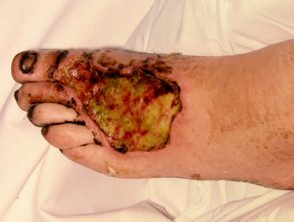
Necrotizing skin infection
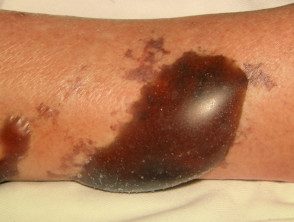
Necrotizing skin infection
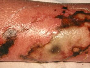
Necrotizing skin infection
Research in necrotizing infections.
Lab tests
Blood cultures should be performed to identify specific organisms involved in the infection.
The table below was developed as a risk indicator for necrotizing fasciitis by Wong et al. in 2004. Scores ≥ 6 were found to have a positive predictive value of 92% and a negative predictive value of 96%.
| laboratory marker | Score evaluation |
|---|---|
| CRP (mg/dL) | <150 > > 150 = 4 points |
| WBC (per mm3) | <15 > 15-25 = 1 point > 25 = 2 points |
| Hb (g/dL) | > 13.5 = 0 points 11.6–13.5 = 1 point <11.5 = 2 puntos |
| Serum sodium | > 135 = 0 points <135 > |
| serum creatinine | ≤ 1.6 = 0 points > 1.6 = 2 points |
| Serum glucose | <180 > > 180 = 1 point |
PCR: C-reactive protein; Half pension: hemoglobin.
Image
Imaging has a limited role in the diagnosis of infectious gangrene.
- Plain radiography can demonstrate subcutaneous emphysema (gas under the skin) sometimes present in clostridial infections.
- Connecticut scan may show fascial thickening, edema, subcutaneous gas and abscess formation.
- Magnetic resonance The scan shows similar findings to CT but is less specific as tissue enhancement can mimic other inflammatory processes.
Surgical exploration
- Surgery is the gold standard for diagnosing necrotizing fasciitis: fascia It separates from the surrounding tissue.
- Little incisions with local exploration are useful to identify necrotic tissue in other cases.
What is the treatment for established wet gangrene?
Treatment for gangrene depends on the location and cause, but focuses on:
- radical surgical debridement +/- amputation
- Spread spectrum antimicrobial Therapy: Penicillin G, clindamycin, vancomycin, and gentamicin can be used for empiric therapy for methicillin-resistant Staphylococcus aureus (MRSA), gram-negative, gram-positive, and anaerobic organisms
- Hemodynamic support.
Administration may also include:
-
Worm debridement therapy: Worms consume dead and infected tissue
- Vascular surgery to improve circulation.
- Hyperbaric oxygen, especially in patients with diabetes or peripheral vascular disease.
-
Intravenous immunoglobulin – This can limit the inflammatory response by binding staphylococcal and streptococcal toxins.
- Treatment of underlying conditions such as diabetes.
the incidence of wartime gas gangrene has been markedly reduced with case identification and isolation, and early debridement, amputation, and supportive treatment.
Which is the forecast for wet gangrene?
The prognosis in wet gangrene depends on the extent of the disease, the underlying cause, and the timing of appropriate treatment.
Anaya et.al have constructed a clinical score to predict patient outcomes for infectious gangrene based on several clinical criteria:
- Heart rate > 110 beats per minute
- Temperature <36 grados centígrados
- Serum creatinine > 1.5 mg/dL
- Age > 50 years
- WBC > 40,000 per uL
- Hematocrit > 50%
Patients with few of these findings had a risk of death of 6% in this study, while patients with several findings had a risk of death of up to 88%. The prognosis can vary widely due to the aggressive nature of gangrene if early treatment is not accessed.
