
What is it bullous systemic lupus erythematosus?
Bullous systemic lupus erythematosus (SLE) is a autoimmune Subepidermal blistering disease that occurs in patients with SLE. It is associated with antibodies against type VII collagen.
Bullous LES is also called bullous eruption of SLE and vesiculobullous SLE
Bullous systemic lupus erythematosus
bullous LES
Who gets bullous systemic lupus erythematosus?
the incidence of bullous SLE was estimated at 0.22 and 0.26 cases per million per year in France and Singapore. In a large cohort of sera taken from patients with immunobullous disorders, 1–2% were identified as bullous SLE [1,2].
Like SLE, bullous SLE usually occurs in women of African descent in their thirties. However, it can occur in all ages, sexes, and ethnicities.
What causes bullous systemic lupus erythematosus?
Bullous SLE is classified into type 1 and type 2.
- Most patients have type I bullous SLE, where autoantibodies are directed against type VII collagen, specificallycollagen type 1 and type 2 domains (NC1 and NC2).
- The NC1 domain plays an important role in maintaining the structure of the dermal–epidermal union (DEJ) [3–5].
- In bullous SLE type II, antibodies are directed against other antigens of the DEJ including BP130, BP280 and plate 332.
Predisposing factors include Adverse drug reactions a hydralazine, penicillamine, methimazole; and ultraviolet exposure (UV) B radiation.
Bullous SLE is immunogenically related to epidermolysis acista bullosa (EBA). In both, the expression of autoimmunity to write collagen VII is associated with humans White blood cell antigen class II allele [6].
What are the clinical characteristics of bullous systemic lupus erythematosus?
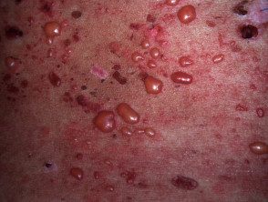
In noisy, tense LES vesicles, bullas and erosions arise in normal or erythematous skin, generally in places exposed to the sun.
- The face is often affected; mucous membrane Surfaces may also be involved.
- The onset of the bullous rash does not correlate with systemic disease activity.
- The blisters may be clear or hemorrhagic contents.
- The blisters heal without scarring.
- Pruritus is absent or mild.
- Milia They are not a standout feature.
- Erythematous cancel urticarial plates may arise.
What are the complications of bullous systemic lupus erythematosus?
No complications have been described.
How is bullous systemic lupus erythematosus diagnosed?
Most patients with bullous SLE have already been diagnosed with SLE. Investigations are necessary to distinguish bullous SLE from SLE with a concomitant blistering disorder Rarely, bullous SLE may be the initial clinical manifestation of SLE.
Histopathological the findings look similar dermatitis herpetiformis, since both have:
- Subepidermal division
- A neutrophils predominant infiltrate on top dermis
- Papillary micro abscesses
- Leukocytoclasis
- mucin statement in the dermis
Basal cap vacuolation, characteristic of other forms of cutaneous LE, is not present.
The direct characteristic immunofluorescence (DIF) find in a perilesional skin biopsy in bulloso LES is linear or granular immunoglobulin (Ig) G, IgM, IgA or C3 at the dermoepidermal junction (DEJ).
Serum autoantibodies, detected by the indirect immunofluorescence test or enzymeLinked immunosorbent assay (ELISA) against collagen type VII (bullous SLE type I). BP 230, BP 180 or lamina 322 can also be targeted (bullous LLE type II).
DIF Sawing Pattern Analysis microscopy differentiates tissue-bound autoantibodies against type VII collagen, where a U-shaped serrated pattern is seen, from all other anti-DEJ antibodies where a serrated pattern is seen [7].
The diagnostic criteria for bullous SLE require:
- A diagnosis of SLE based on the American College of Rheumatology criteria
- An acquired vesiculobular rash
- Histopathology evidence of a subepidermal blister and a neutrophil-dominant dermal infiltrate
-
DIF microscopy showing linear or granular deposits of immunoglobulin (Ig) G or IgM and often IgA in the basement membrane area
- Antibodies against collagen type VII by indirect immunofluorescence in salt-split skin or ELISA.
Which is the differential diagnosis for bullous systemic lupus erythematosus?
Other blistering conditions that may resemble bullous SLE include:
-
Bullous drug eruption: Band-like deposits of IgG, IgM, and IgA may be present; however, no linear or granular deposits are observed.
- The pemphigoid group of diseases: bullous pemphigoid, linear IgA disease, epidermolysis bullosa and dermatitis herpetiformis can be differentiated for the absence of SLE, according to the criteria of the American College of Rheumatology
- Skin lesions of highly active SLE: blisters may rarely form; A skin biopsy will confirm extensive vacuolation in the DEJ.
What is the treatment for bullous systemic lupus erythematosus?
Dapsone, at a dose of 1.0 to 1.5 mg/kg/day, is considered the treatment of choice. It is effective in the vast majority of patients and leads to rapid clinical improvement within a few days to a few weeks. This rapid response differentiates bullous SLE from SLE with a concomitant autoimmune bullous disease.
When dapsone fails, prednisolone (systemic steroids) and azathioprine are used. Patients successfully treated with methotrexate or rituximab have been reported [8–10].
What is the outcome of bullous systemic lupus erythematosus?
Bullous SLE is transient in most cases and usually regresses completely without further flares, regardless of systemic disease activity.
