What is it eczema herpeticum?
Eczema herpeticum is a disseminated viral infection characterized by fever and clusters of itchy or perforated blisters erosions. It is most often seen as a complication of atopic dermatitis/eczema.
Eczema herpeticum is also known as Kaposi varicelliformis. eruption because it was initially described by Kaposi in 1887, who thought it resembled chickenpox/varicella.
What is the cause of eczema herpeticum?
Most cases of eczema herpeticum are due to Herpes Simplex of type 1 or 2.
Eczema herpeticum usually appears during a first episode of infection with Herpes simplex (primary herpes). Signs appear 5 to 12 days after contact with an infected person, who may or may not have a visible cold sore.
Eczema herpeticum can also complicate recurrent herpes. However, repeated episodes of eczema herpeticum are unusual.
Eczema herpeticum can affect men and women of all ages, but is most often seen in infants and children with atopic dermatitis. Patients with atopic dermatitis appear to have reduced immunity to herpes infection. Your underlying dermatitis may be mild to severe, active or inactive.
Eczema herpeticum is better called Kaposi's varicelliform eruption when a breakdown of the skin barrier is not due to eczema. Examples of noteczematous conditions prone to severe located Herpes infections are:
- Thermal burns
- Pemphigus vulgaris
- Darier's disease
- Benign family pemphigus
- Cutaneous T cell lymphoma/ mycosis fungoides
- Ichthyosis.
Other viruses may occasionally be responsible for a similar rash, such as eczema coxsackium due to coxsackievirus A16 (the cause of hand, foot, and mouth disease).
Since smallpox has been eliminated, the vaccine disseminated as a consequence of the smallpox vaccine is now very rare. It was reported to be very serious, with mortality up to 50%.
What are the clinical features of eczema herpeticum?
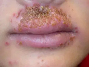
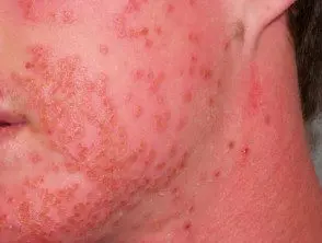
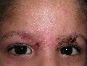
Eczema herpeticum begins with clusters of itchy, painful blisters. It can affect any site, but is most often seen on the face and neck. Blisters can occur on normal skin or on sites actively or previously affected by atopic dermatitis or other skin disease. New patches form and spread over 7 to 10 days, rarely spreading widely throughout the body.
The patient is unwell, has fever and local swelling. lymph nodes.
- The blisters are monomorphic, that is, they all appear similar to each other.
- May be filled with pale yellow or thick fluid purulent material.
- They are often stained with blood, that is, red, purple or black.
- New blisters have central dimples (umbilication).
- They may cry or bleed.
- old blisters Cortex on and form sores (erosions)
- Lesions heal in 2 to 6 weeks.
- In severe cases where the skin has been destroyed by infection, small white scars can persist long-term.
Secondary bacterial Staph or strep infection can lead to impetigo and cellulitis.
Severe eczema herpeticum can affect multiple organs, including the eyes, brain, lungs, and liver. It can rarely be fatal.
Herpetic eczema

Herpetic eczema

Herpetic eczema

Herpetic eczema
See more images of eczema herpeticum...
How is eczema herpeticum diagnosed?
Eczema herpeticum can be diagnosed clinically when a patient with known atopic dermatitis has a acute painful monomorphic clustering eruption vesicles associated with fever and discomfort. Viral infection can be confirmed with viral swabs taken by scraping the base of a fresh blister. Various laboratory tests are available.
- Viral culture
- direct fluorescent antibody stain
- PCR (Polymerase chain reaction) sequencing
-
Tzank spot showing epithelial multinucleated giant cells and acantholysis (cell separation)
Bacterial swabs should also be taken to microscopy and culture as eczema herpeticum may resemble impetigo and may be complicated by secondary bacterial infection.
Skin biopsy reveals distinctive pathological changes.
What is the treatment of eczema herpeticum?
Eczema herpeticum is considered one of the few dermatological emergencies. timely treatment with antiviral the medication should eliminate the need for hospital admission.
Oral acyclovir 400–800 mg 5 times a day, or, if available, valacyclovir 1 g twice a day, for 10–14 days or until lesions heal. Intravenous acyclovir is prescribed if the patient is too ill to take tablets, or if the infection deteriorates despite treatment.
Secondary bacterial infection of the skin is treated with systemic antibiotics
Current Steroids are generally not recommended but may be necessary to treat active atopic dermatitis.
consult a ophthalmologist when eyelid or eye involvement is seen or suspected.
