What is it linear focal elastosis?
Elastosis refers to abnormal or increased statement of elastin fibers within the dermis.
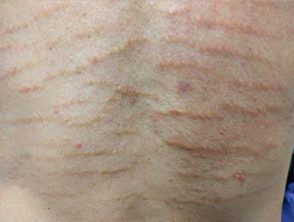
Focal linear elastosis is a rare form of dermal elastosis that resembles stretch marks.
Linear focal elastosis

Who gets focal linear elastosis?
There are few case reports of focal linear elastosis, in part because it can be confused with more common stretch marks. The truth predominance of focal elastosis is unknown [1].
More cases of focal linear elastosis have been reported in men than in women. The average age range of onset is adolescence and ranges between 7 and 89 years. [1,2]. There is no race or ethnicity predilection [3]. Strong hereditary the factors have not been identified [1].
What causes focal linear elastosis?
the Pathogenesis of focal linear elastosis is uncertain.
Focal linear elastosis and stretch mark distension can coexist in the same distribution in affected individuals [3]. Focal linear elastosis may represent excessive regeneration of damage elastic fibers as part of the repair process of striae distensae [6]. However, the clinic and histopathological The appearances of linear focal elastosis and striae distensae differ.
No clear triggering factor for linear elastosis has been identified, although compelling trauma, pregnancy, weight loss or rapid growth may be involved [1].
What are the clinical characteristics of focal linear elastosis?
Linear focal elastosis appears yellow, palpable lines that extend horizontally over the lower back. There are also reports of focal linear elastosis on the trunk, lower extremities, and face. [4,5].
Focal linear elastosis is usually asymptomatic and is diagnosed incidentally. Not known to be associated with systemic disease.
Which is the differential diagnosis for focal linear elastosis?
The main differential diagnosis for focal linear elastosis is stria distensae. However, stretch marks are usually red or white and depressed to palpation.
How is focal linear elastosis diagnosed?
Focal linear elastosis is diagnosed clinically by its appearance, distribution, and skin. biopsy.
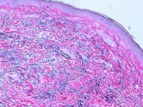
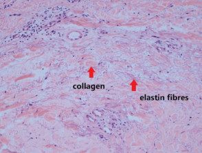
Histology demonstrates increased elastic fibers within the middle dermis. The fibers appear as elongated material that separates the dermal skin. collagen. Elastic tissue staining may be useful to appreciate excess elastin. Early lesions can paradoxically show elastolysis (destruction of elastin fibers) [1].
Histology of linear focal elastosis

Low power view. Elastin stain

High power view
How is focal linear elastosis treated?
No effective treatment has been described for focal linear elastosis. Current steroid and topical retinoid have not shown any benefit [1].
