Category

Skin Tumors

Tumor removal with aesthetic results

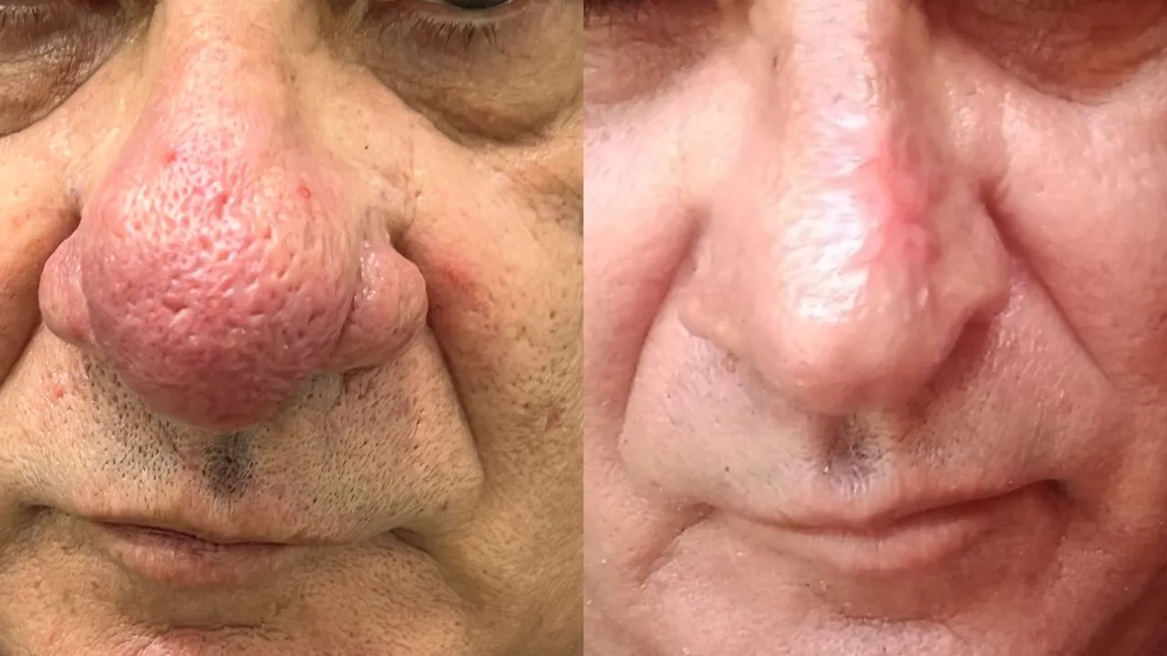
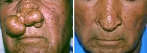
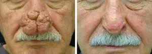
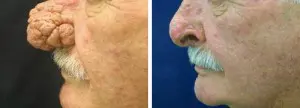
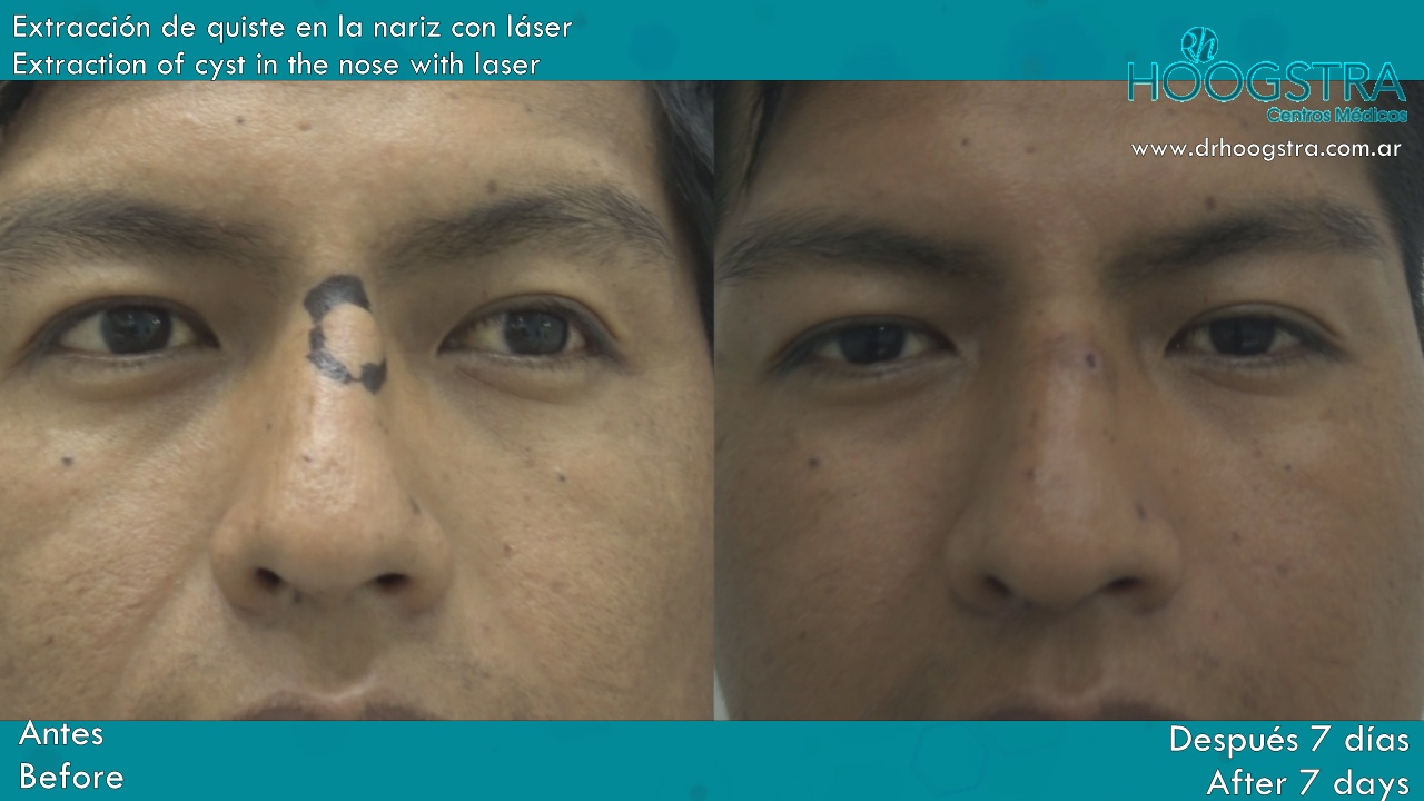
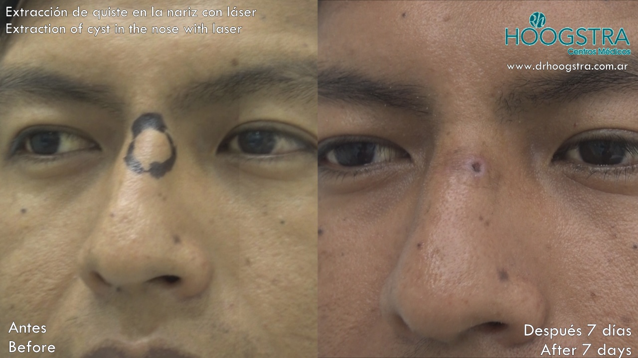
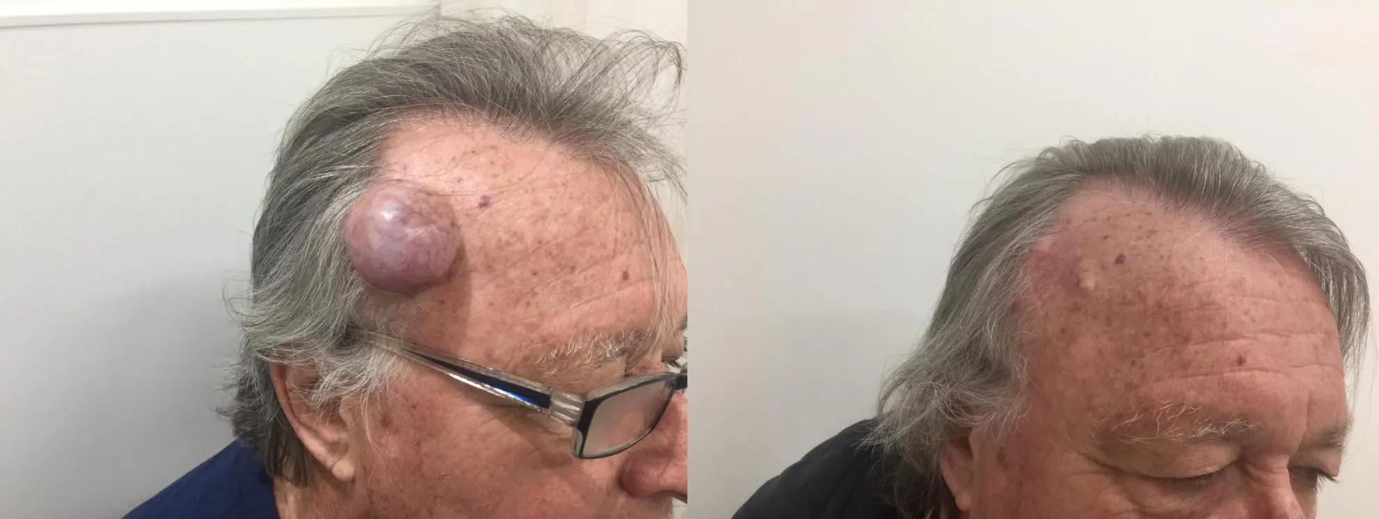
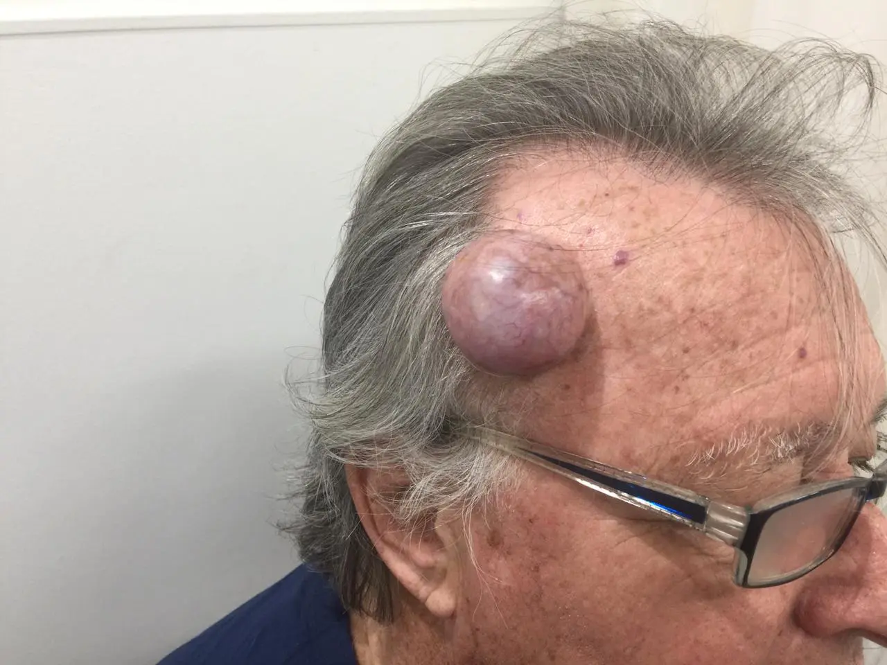
Es la extracción quirúrgica del tumor generando la menor cicatriz posible pero asegurando la extracción total de la lesión para luego, mediante técnicas de reconstrucción, permitir un resultado estético y funcional óptimo.

¿En qué casos se recomienda?

Está indicada en todos los casos, siendo necesario el estudio anatomopatológico (biopsia) del tumor para confirmar el diagnóstico y la resección total de la lesión.

¿Cómo se realiza?

El procedimiento se realiza bajo anestesia local y en algunos casos bajo sedación. En algunos casos es necesario recurrir a la extracción de injertos de tejidos para la correcta reconstrucción del párpado. No existen riesgos relevantes a tener en cuenta.

Doubts and queries

If you have any medical questions, send an email to [email protected]  . In this mail Only medical consultations will be received. Inquiries about shifts, payments and administrative issues are not attended.

By phone on Monday, Thursday and Friday from 8 a.m. to 3 p.m. at the following telephone numbers.

  • 0054 11 4 901 6690
  • 0054 11 4 904 3434
  • 0054 11 4 904 0880

Schedule consultation

Book a consultation at our medical centers located in Caballito, Belgrano and Pilar


Siliconoma


Siliconoma is a foreign body granulomatous reaction that occurs as a result of the silicone subcutaneous implant, generally for cosmetic purposes.

Clinical picture

The siliconoma (s) appear months to years later, after application. Treatment of this condition is difficult since the results obtained are incomplete and transitory.
Liquid silicone was considered useful as a filler material because it is permanent and minimally antigenic, but its use in injectable form has not been approved by any health authority because it migrates, presents granulomas, damages tissues, causing severe inflammatory reactions, and is very difficult to extract. of the organism.

For many years, at Hoogstra Medical Centers we have seen the unpleasant aesthetic effects and the damage to the health and psyche of patients who have received this type of practice poorly informed.

Treatment

We have carried out all kinds of procedures to help affected patients and we have currently discovered that some well-capped siliconomas can be removed effectively, quickly and safely through micro-perforations produced in the skin with the Lotus II laser.

As we see in this video, the siliconoma was completely drained with the use of the Lotus laser, no previous preparation was required, no anesthesia was used and the patient was able to continue immediately with his daily activity, with a cure of 100%.

We do not consider this procedure to be effective in all affected patients, but in many it will be an effective solution without additional tissue and patient damage.

Doubts and queries

If you have any medical questions, send an email to [email protected]  . In this mail Only medical consultations will be received. Inquiries about shifts, payments and administrative issues are not attended.

By phone on Monday, Thursday and Friday from 8 a.m. to 3 p.m. at the following telephone numbers.

  • 0054 11 4 901 6690
  • 0054 11 4 904 3434
  • 0054 11 4 904 0880

Schedule consultation

Book a consultation at our medical centers located in Caballito, Belgrano and Pilar

Rhinophyma


Rhinophyma is clinically characterized by an increase in the size of the nose, a process practically exclusive to adult men.

Clinical picture

The surface of the nose shows a cobblestone appearance due to the prominence of multiple sebaceous glands; there is also dilation of the excretory pores with spontaneous seborrhea and pressure.

Etiopathogenesis. Rhinophyma is a variety of rosacea in which the growth of sebaceous glands is stimulated.

Treatment

Conventional treatments are surgery, cryotherapy and electrocoagulation. Currently, the most effective treatment with the best aesthetic results is that obtained with Carbon Dioxide Laser used in our Medical Centers.

Doubts and queries

If you have any medical questions, send an email to [email protected]  . In this mail Only medical consultations will be received. Inquiries about shifts, payments and administrative issues are not attended.

By phone on Monday, Thursday and Friday from 8 a.m. to 3 p.m. at the following telephone numbers.

  • 0054 11 4 901 6690
  • 0054 11 4 904 3434
  • 0054 11 4 904 0880

Schedule consultation

Book a consultation at our medical centers located in Caballito, Belgrano and Pilar

Eruptive hairy cysts


Clinical picture

El quiste velloso eruptivo es un trastorno en el desarrollo del folículo piloso, el cual conduce a la oclusión del infundíbulo produciendo retención de pelos y dilatación quística.

Se manifiesta clínicamente por la aparición de numerosas máculas o pápulas asintomáticas en cara anterior de tórax, abdomen y axilas, aunque también pueden presentarse en cuello, ingles y rostro.

Las lesiones pueden ser del color de la piel, eritematosas, amarillentas o azuladas; su tamaño varía de 1-5 mm; y la cantidad es variable.

Treatment

El tratamiento de estos quistes hasta la actualidad era difícil. Hoy en día gracias a la tecnología Láser se pueden realizar pequeñas perforaciones en la piel, totalmente indoloras y drenar el contenido del quiste obteniendo excelentes resultados.

Patient testimonial

We invite you to watch the following videos with the story of patients who have received this treatment in our Medical Centers

Doubts and queries

If you have any medical questions, send an email to [email protected]  .

Inquiries can also be made through WhatsApp in the number +54 9 11 5503-5126 

By phone on Monday, Thursday and Friday from 8 a.m. to 3 p.m. at the following telephone numbers.

  • 0054 11 4 901 6690
  • 0054 11 4 904 3434
  • 0054 11 4 904 0880

Schedule consultation

Book a consultation at our medical centers located in Caballito, Belgrano and Pilar

Dermoid cysts


Los quistes dermoides se consideran una forma de teratoma quístico derivado del epitelio germinal embrionario. Frecuentemente en el interior del quiste encontramos piel, pelo, glándulas, dientes, etc.

Etiología

Cuando las líneas óseas de unión comienzan a cerrarse, puede quedar tejido epitelial atrapado en las mismas. Este tejido atrapado es el que forma el quiste.

Las suturas óseas más frecuentemente afectadas son las frontozygomaticas, esto da origen a que el quiste se localice con más frecuencia en la cola de la ceja.

Otras localizaciones de los quistes dermoides son los ovarios y la columna vertebral.

Clinical picture

Los Quistes Dermoides son masas ovales, indoloras, lisas, de crecimiento lento.

Differential diagnosis

Si el quiste se localizara en la región media puede ser un encefalocele.

Treatment

El tratamiento es quirúrgico y consiste en la exéresis completa de la lesión.
Es más sencillo remover completamente el quiste y no dejar cicatrices utilizando el Láser de Dióxido de carbono.
Si el quiste se infectó y se derramó su contenido es más probable que en un futuro tengamos una recidiva del mismo.
Otras causas de recidiva se presentan cuando el quiste se mete en forma profunda en la sutura ósea y esto dificulta su extirpación completa.

Doubts and queries

If you have any medical questions, send an email to [email protected]  .

Inquiries can also be made through WhatsApp in the number +54 9 11 5503-5126 

By phone on Monday, Thursday and Friday from 8 a.m. to 3 p.m. at the following telephone numbers.

  • 0054 11 4 901 6690
  • 0054 11 4 904 3434
  • 0054 11 4 904 0880

Schedule consultation

Book a consultation at our medical centers located in Caballito, Belgrano and Pilar