
What is amyloidosis?
Amyloidosis is a group of disorders in which amyloid The protein is deposited in various tissues. It includes:
- Systemic amyloidosis, which involves a variety of organs
- Primary cutaneous amyloidosis in which only the skin is affected.
What is amyloidosis? skin dyschromic?
Amyloidosis cutis dyschromica is a form of primary cutaneous amyloidosis that causes located hyperpigmentation and hypopigmentation.
Amyloidosis cutis dyschromica was first described by the Japanese dermatologist Morishima in 1970 [1].
Amyloidosis cutis dyschromica *
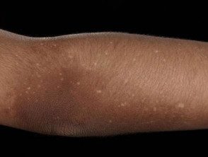
Figure 1. Multiple dotted areas of hypopigmentation on a background of irregular hyperpigmentation on
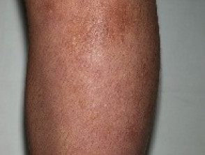
Figure 2. Multiple subtle areas of hypopigmentation points on a background of irregular hyperpigmentation
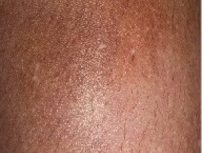
Figure 3. A close-up image of the pigment change in the lower leg of a patient affected by amyl.
Who gets Amyloidosis cutis dyschromica?
Amyloidosis cutis dyschromica is rare. As of this writing, around 50 cases have been reported in the medical literature. [2].
- Men and women are equally affected.
- Most patients develop skin first. pigment changes in early childhood.
- The majority of patients (77%) have a family history of similar pigmentary abnormality.
- Most cases (63%) have occurred in ethnic East Asian (Chinese, Hong Kong Chinese, and Taiwanese) or Southeast Asian (Thai, Filipino, and Indonesian) ethnicity.
- Amyloidosis cutis dyschromica is also commonly seen in people of Indian and Pakistani descent.
- Only three cases of cutis dyschromic amyloidosis have been reported in Caucasian patients.
What causes amyloidosis cutis dyschromica?
Amyloidosis cutis dyschromica is caused by statement amyloid derived from degenerated or damaged keratinocytes. The reason why keratinocytes degenerate and how this relates to the loss and gain of pigment in the affected areas of the skin is unclear. A genetic the cause is probable
What are the clinical features of cutis dyschromica amyloidosis?
Amyloidosis cutis dyschromica slowly causes progressive localized hyperpigmentation and hypopigmentation (dyschromic or dyschromatosis).
- In most cases, the onset of pigment change is in childhood, although in some individuals the changes are not noticeable until adulthood.
- Dyschromic does not affect palms, soles and mucous membrane surfaces
- Facial involvement is rare (approximately 10%) [2].
The specific findings that distinguish amyloidosis cutis dyschromica from the more common macular and lichenoid Variants of primary cutaneous amyloidosis are:
- Dotted, lattice or diffuse hyperpigmentation mixed with "lentil size" hypopigmented macules.
- Mild or not associated pruritus.
What are the complications of amyloidosis cutis dyschromica?
There are no known complications of amyloidosis cutis dyschromica.
How is amyloidosis cutis dyschromica diagnosed?
Amyloidosis cutis dyschromica is diagnosed by recognizing the typical clinical features and confirmed by the biopsy of a hyperpigmented or hypopigmented taint.
-
- Amyloid deposits are present on the papilla. dermis just below the epidermis.
- They can be very subtle and difficult to see with hematoxylin and eosin (H&E) staining, thus requiring Congo red staining.
- Using polarized light, the apple-green birefringence of amyloid material can be seen in microscopic exam.
Histology of amyloidosis cutis dyschromica *
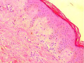
Figure 4. A skin biopsy showing very subtle pink blood cells just below the epidermis in the amyloids.
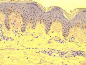
Figure 5. A biopsy of skin stained with Congo red staining, highlighting the observed amyloid deposits
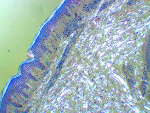
Figure 6. A skin biopsy Sample stained Congo red seen under polarized light microscopy showing a
Which is the differential diagnosis for amyloidosis cutis dyschromica?
Amyloidosis cutis dyschromica can closely resemble other rare dyschromatoses; These include:
- Hereditary universal dyschromatosis
- Hereditary symmetric dyschromatosis
- Tricolor complexion
-
Simple epidermolysis bullosa with mottling pigmentation
- Tertiary paint.
Other conditions that should be excluded include:
- Dyskeratosis congenita, presented with a nail dystrophy, oral leukoplakia and bone marrow failure in childhood
-
Xeroderma pigmentosum, which causes skin marking and ocular photosensitivity and aggressive skin cancers beginning in childhood
-
Degos disease, a life-threatening disease vasculopathy.
What is the treatment for amyloidosis cutis dyschromica?
Not therapeutic Intervention has been shown to be beneficial in cutis dyschromica amyloidosis. Multiple current treatments, including urea to 10% cream and tazarotene [1], have been prescribed to patients without significant improvement. Oral supplements of vitamin C and vitamin E have had minimal benefit [3]. Acitretin has been prescribed in a small number of cases, with a modest improvement reported in approximately 85% of cases. [2].
What is the result of amyloidosis cutis dyschromica?
Amyloidosis cutis dyschromica is typically gradually progressive. Dyschromia can affect almost all of the skin, except the palms, soles of the feet, and the oral and genital tract. mucous membrane. Alternatively, amyloidosis cutis dyschromica can progress gradually in localized areas, and is often more prominent in areas of skin that lie on bony prominences and joint surfaces.
