
What is cellulite?
Cellulite is a acute bacterial infection of the dermis and subcutaneous skin tissues. It is commonly caused by anyone Streptococcus pyogenes or Staphylococcus aureus. Cellulite presents as an enlarged area of red, warm, swollen and tender skin. Cellulite can occur anywhere on the body, but is most often seen on the lower leg. May be accompanied by lymphangitis, which appears as a red streak after lymphatic ships to local lymph node (which can also be tender and enlarged).
The signs of cellulite are nonspecific and the diagnosis is based primarily on the patient's medical history and physical examination. A number of skin conditions can "mimic" cellulite, and about 30% of cellulite cases are misdiagnosed. Distinguishing between cellulite and these simulating conditions is important to avoid unnecessary treatments and complications, and to speed up proper treatment.
Cellulitis
What are the clinical characteristics of cellulite?
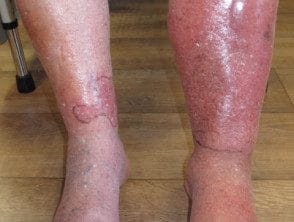
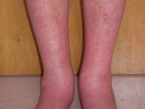
Unilateral distribution in the lower leg
Cellulite of the legs is almost always unilateral. Bilateral Cellulite distribution rarely occurs, usually as a result of an underlying condition, such as lymphedema. The bilateral distribution of a eruption In the absence of other symptoms of cellulite, an alternative diagnosis should be sought.
Response to treatment
If not treated, cellulite will progress rapidly. If appropriate antibiotics are started, cellulite should respond within hours to days. Lack of response suggests an alternative diagnosis.
Symptoms and signs of infection.
A cellulite patient may experience symptoms such as nausea and discomfort.
Signs of cellulite infection include:
- A feeling of intense heat in the affected area.
- Fever
- Tachycardia
- Leukocytosis
- High inflammatory markers (for example, C-reactive protein [[CRP], erythrocytes sedimentation rate [[ESR])
Infection risk factors.
There may be an entry portal, such as a ulcer or injury, which allows bacteria to break the skin's defenses. Other risk factors include chronic edema, lymphedema, obesity and diabetes mellitus.
What are the clinical conditions that commonly mimic cellulite?
Other cutaneous infections
Other skin infections that share characteristics similar to cellulite are erysipelas, necrotizing fasciitis and Herpes zoster.
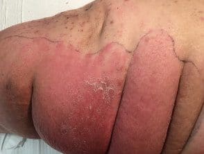
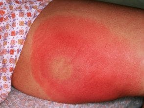
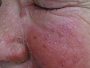
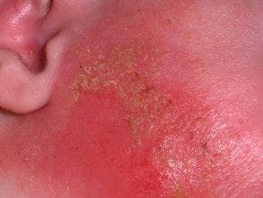
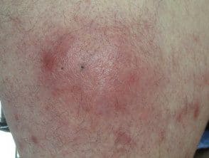
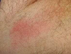
Erysipelas
Erysipelas is a superficial variant of cellulite that is also predominantly caused by S. pyogenes or S. aureus.
Erysipelas occurs with:
- Abruptly demarcated erythema
- Burning
- Edema
- Intense heat of the affected area
- Systemic symptoms, such as fever, malaise, and nausea.
Erysipelas
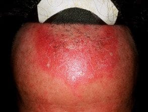
Necrotizing fasciitis
Necrotizing fasciitis is a bacterial infection that affects the subcutaneous tissues. As it does not affect the epidermis and upper dermis, its characteristics may be subtle at the initial stage, but infection later results in rapid and severe tissue necrosis if not treated Most patients with necrotizing fasciitis are seriously ill with septic features.
The keys to diagnosing necrotizing fasciitis include:
- Severe pain, apparently out of proportion to clinical findings.
- Edema or tenderness that extends beyond the erythematous edge of affected area
- Cutaneous gangrene and blisters
- Crepitation (due to the subcutaneous gas produced by anaerobic organisms)
- Fluctuation, indicating purulent material in the soft tissues
- Rapid expansion despite antibiotic therapy.
Necrotizing fasciitis
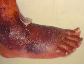
Necrotizing fasciitis
Necrotizing fasciitis
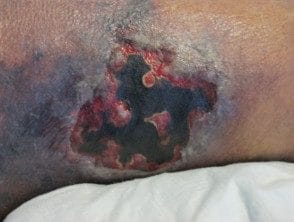
Necrotizing fasciitis
Herpes infection
Some of the symptoms of herpes zoster (shingles) are similar to cellulite, including:
- Sudden onset of fever
- Located rash with pain
- Swelling
- Hot
- Redness
However, herpes zoster is characterized by its dermatomal distribution and umbilical vesicles. Antibiotics are not effective against the herpes zoster virus, which is treated with antiviral agents, such as acyclovir.
Herpes infection
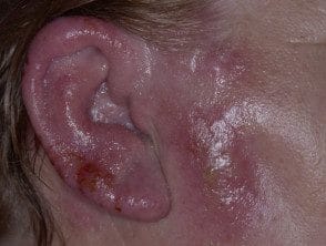
Herpes infection
Herpes infection
Herpes infection
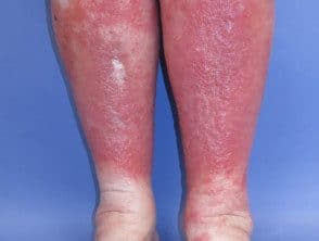
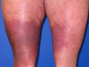
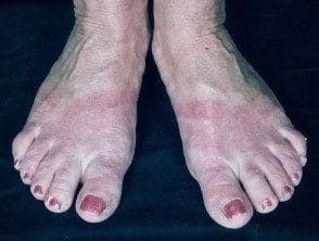
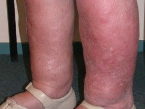
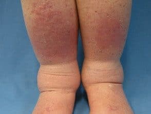
Venous disease
Venous disease of the lower legs affects about 20% in people over the age of 70. Damage or degeneration of leg veins. It is a very common reason for red legs. Signs of venous disease can include:
- Swelling due to accumulation of blood.
- Color changes
- Dry and peeling skin
- Firm hardening (described as a “tied” appearance)
- Swelling of the calves over the narrow ankles (sometimes described as an 'inverted champagne bottle' appearance).
These changes have been classified within a framework of symptoms known as clinical, etiological, anatomical and pathophysiological classification (CEAP).
Dependent blush
In this condition, the dilated vessels of the skin cause a red and dark discoloration of the legs when sitting or standing. Patients are typically asymptomatic.
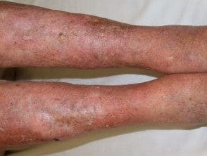
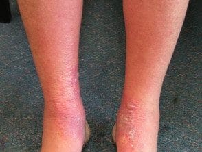
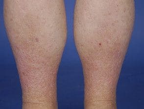
Venous eczema
Prolonged edema leads to inflammation and subsequent redness, itching, dryness and climbing and what is known as venous eczema (also called stasis dermatitis). Venous eczema is usually not as red, hot, or edematous like cellulite
Venous eczema
Venous eczema
Venous eczema
Venous eczema
Venous thrombosis
Superficial thrombophlebitis causes redness, warmth, and cellulite-like sensitivity, except that the inflammation is linear, following the course of the underlying thrombosed vein, which is palpable and tender The edema is absent and the skin is not taut, smooth or shiny. There are no systemic symptoms.
The inflammation of deep vein thrombosis does not spread to the skin surface or cause erythema.
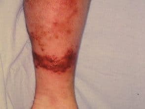
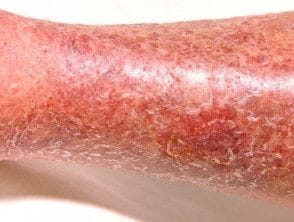
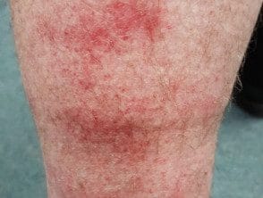
Lipodermatosclerosis
Lipodermatosclerosis can be acute or chronic. Acute lipodermatosclerosis is often tender and bright red in color, and occurs with or without edema. Systemic symptoms and signs of infection are absent. Later, a reddish brown discoloration indicates prolonged inflammation and statement of hemosmosin (an insoluble form of iron that has leaked from inflammation capillaries)
Chronic lipodermatosclerosis has a “tied” appearance due to fibrosis (hburn) from the deeper tissues.
Lipodermatosclerosis
Acute lipodermatosclerosis
Acute lipodermatosclerosis
Chronic lipodermatosclerosis
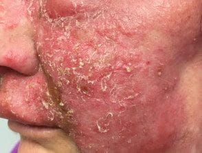
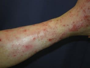
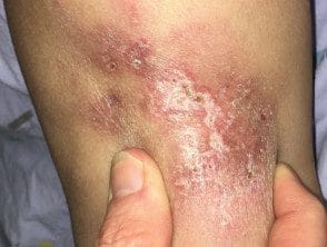
Dermatitis
Dermatitis or eczema describes a group of common inflammatory skin conditions. Leg dermatitis may be due to atopic dermatitis, discoid eczema, contact dermatitis, craquelé eczema (due to dry skin) or venous eczema. Combinations may occur.
Acute eczema
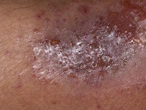
Acute eczema can mimic cellulite, regardless of the cause; It is characterized by erythema, crying, crust, and blisters and may be secondarily infected (impetiginization)
Acute eczema
Acute eczema
Acute eczema
Acute eczema
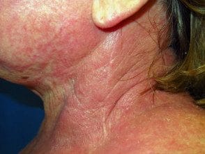
Contact dermatitis
Contact dermatitis is classified as contact irritating dermatitis or allergic contact dermatitis (a type IV hypersensitivity reaction).
Irritants Those that cause contact dermatitis include soaps and detergents.
Potential Allergens Which can cause contact dermatitis include:
-
Rosin in adhesive bandages
-
Rubber accelerators in elastic bandages.
-
Preservatives in moisturizers and current medicines
Contact dermatitis presents with blisters or scalyerythematous plates, What are they often irregularly and clearly delimited. Patients may have a history of exposure to a relevant irritant or allergen. Symptoms and systemic signs are absent.
Contact dermatitis
Contact dermatitis
Contact dermatitis
Contact dermatitis
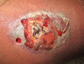
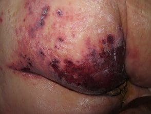
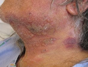
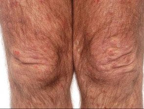
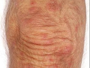
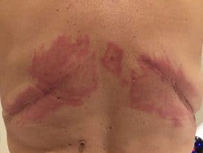
Panniculitis
Panniculitis refers to a group of conditions in which there is subcutaneous inflammation of the fat. Panniculitis is characterized by red, firm, tender plaques or nodules who has a predilection for the lower legs
The most common form of panniculitis is erythema nodosum, which can be distinguished from cellulite by its bilateral and multifocal distribution. recurrent nature and absence of edema. Patients may have systemic symptoms, either from an underlying cause or from panniculitis itself, which can cause fever, malaise, and arthralgia.
Panniculitis
Lupus panniculitis
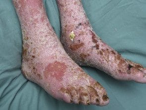
Lymphedema
Lymphedema is a common mimic of cellulite. The underlying Pathogenesis Lymphedema is similar to venous disease, in which there is a decrease in tissue oxygenation as a result of extravasated lymph.
The clinical features of lymphedema include:
- Not for a long timebites edema with induration
- Erythema
- Hyperkeratosis
- Like a viral wart papules and plates.
the inguinal lymphatic it may have been damaged or occluded for filariasis, lymph node dissection, radiotherapy, or obesity. Infectious signs and symptoms are absent.
Lymphedema is associated with a risk of skin infection, including cellulite, due to impaired lymphatic drainage.
Lymphedema
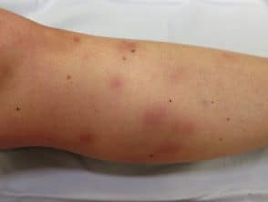
Eosinophilic cellulitis
Eosinophilic cellulitis, or Wells syndromeIt is characterized by recurrent itchy or painful plaques of unknown cause in which eosinophils are on the skin biopsy. A minority of patients with eosinophilic cellulitis may experience discomfort and fever, but multiple lesions and a recurring history should distinguish it from cellulite. The plates are generally bright red at first, then fade for 4–8 weeks, leaving green, gray, or brown spots.
Eosinophilic cellulitis
Wells syndrome
Wells syndrome
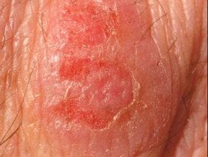
Wells syndrome
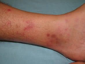
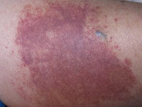
Small container vasculitis
Small vessel vasculitis is characterized by tender, palpable erythematous plaques. purple. Most often it affects both legs. There are various causes
Vasculitis can be accompanied by ulceration, mild blisters and systemic symptoms, such as fever and malaise. Multiple and nopallor Lesions distinguish it from cellulite. The biopsy will generally show leukocytoclastic vasculitis
Small vessel vasculitis
Small vessel vasculitis
Small vessel vasculitis
Small vessel vasculitis
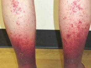
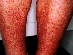
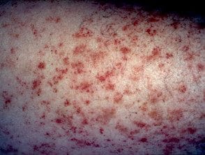
Capillaritis
The capillaritis can be unilateral or bilateral, acute, chronic or recurrent. It favors the lower part of the legs. It is characterized by 'cayenne pepper' petechiaebut these can resemble cellulite when confluent and accompanied by erythematous patches and edema. The leg is cold and there are no systemic symptoms.
Capillaritis
Other cellulite mimics
Rare cellulite mimics include:
- Carcinoma erysipeloid
- Hereditary periodic fever syndromes
-
Erythromelalgia.
Imitates rare cellulite
Erysipeloid carcinoma
Erythromelalgia
Periodic fever
