
What is dermatofibrosarcoma protuberant?
Dermatofibrosarcoma protuberans is a rare skin tumor that arises in the deepest layer of the skin (the dermis) It grows slowly but has a tendency to reappear after excision. Fortunately, it rarely spreads beyond the skin.
Dermatofibrosarcoma protuberans
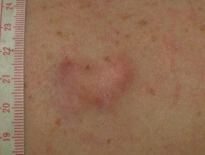
Injury in 2014
Same injury in 2015
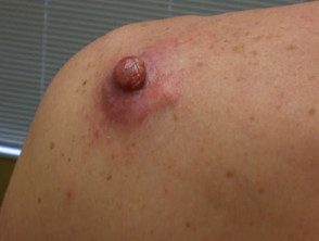
Another example
What causes dermatofibrosarcoma protuberans?
The cause of dermatofibrosarcoma protuberans it is unknown, but an injury to the affected skin may be a predisposing factor. Recent Advances Show Tumor Cells Are Abnormal chromosomes within tumor cells - t (17; 22) (q22; q13) - resulting in fusion gene COL1A1-PDGFB. This encodes a protein that causes a tumor to grow by autocrine overproduction of platelet-derivative growth factor (PDGF).
Who is at risk for dermatofibrosarcoma protuberans?
reermatofibrosarcoma protuberans it is rare and affects less than 1 person per 100,000 residents per year.
- It usually occurs in early to mid adult life between the ages of 20 and 59, but all ages can be affected. The tumor is rare in children.
- Males are affected slightly more often than females.
- There doesn't seem to be any race predilection
What are the signs and symptoms of dermatofibrosarcoma protruding?
reermatofibrosarcoma protuberans usually presents as a painless thickened area of skin (license plate) me nodule that feels rubbery or firm to the touch and sticks to the underlying skin. It can be red-brown or skin-colored. It usually grows very slowly over months or years.
In rare cases, it presents as a soft, depressed area of skin that makes diagnosis even more difficult. reermatofibrosarcoma protuberans it can vary in size from 0.5 to 25 cm in diameter. Fifty to sixty percent of tumors arise on the trunk, often in the shoulder and chest area. The remaining 35% of tumors are found in the extremities and 10 to 15% in the head and neck region.
reermatofibrosarcoma protuberans It is often diagnosed when it enters a more rapid growth phase that results in larger lesions. Neglected tumors can reach large proportions.
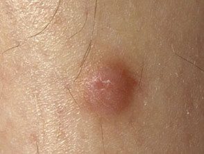
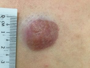
More examples of dermatofibrosarcoma protuberans
Dermatofibrosarcoma protuberans
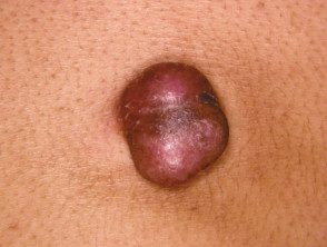
Dermatofibroscarcoma protuberans
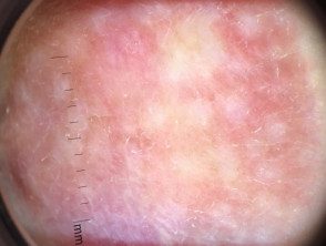
Dermoscopy view
How is dermatofibrosarcoma protuberans diagnosed?
The absence of symptoms often leads to a delay in the diagnosis of reermatofibrosarcoma protuberans. Redness and pain only occur in 15% of cases. It is often confused with other skin conditions, particularly in the early stages. Dermoscopy is not diagnostic as the characteristics of reermatofibrosarcoma protuberans are unspecific Skin biopsy It is necessary to confirm the diagnosis.
Dermatofibrosarcoma protuberans It has a characteristic appearance under the microscope with cells densely arranged in a spindle shape. Complete removal can be difficult to assess due to extensive extensions to the skin and deeper structures. It is important to identify fibrosarcomatous reermatofibrosarcoma protuberans, a more aggressive tumor, which requires more aggressive treatment.
reermatofibrosarcoma protuberans's chromosomal Abnormalities can be detected using reverse transcription.Polymerase chain reaction (RT-PCR) or fluorescence in the place hybridization (FISH)
In most cases of reermatofibrosarcoma protuberans, no further investigations are necessary. If there is suspicion of metastasis or there is a fibrosarcomatous transformation, lymph node ultrasound, chest x-ray and pelvic ultrasound can be arranged.
What is the treatment for dermatofibrosarcoma protuberans?
Treatment of reermatofibrosarcoma protuberans and reermatofibrosarcoma protuberans with fibrosarcomatous transformation consists of wide excision of the injury including deep fascia, with 1–3 cm margin of normal skin. It may take more than one surgical procedure to ensure complete removal of a tumor.
Mohs micrographic surgery, which is a special surgical technique for control tumor margins, sometimes used to verify that all abnormal cells have been removed. Reappearance after Mohs surgery it is reported to be around 1%.
Radiotherapy it is sometimes used in addition to surgery if a tumor cannot be completely removed by surgery. Chemotherapy It is ineffective.
the tyrosine kinase The inhibitory imatinib mesylate is used to treat rare cases of local advanced inoperable or metastatic dermatofibrosarcoma characterized by COL1A1-PDGFB fusion gene, with 50% response rates.
Which is the forecast for dermatofibrosarcoma protuberans?
Follow-up with clinical examination of the reermatofibrosarcoma protuberans It is recommended every 6 months for 5 years, and then annually.
The tumor only metastasizes in 5% of cases. It spreads on 1% via lymphatic vessels to the regional lymphatic glands and at 4% through the bloodstream, most commonly to the lung followed by the brain, bones, and heart.
Local recurrences arise in 11-20% of cases, generally within 3 years of the initial surgery, so follow-up is important. Recurrences are treated surgically as described for the original. primary tumor.
