
What is it gangrene?
Gangrene is the located death of body tissue. Dry gangrene is due to prolonged ischemia (heart attack) or inadequate oxygenation or lack of blood flow. Ischemia affecting proximal blood vessels it usually affects the lower extremities. Ischemia of the peripheries can cause gangrene of the fingers and toes.
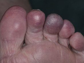
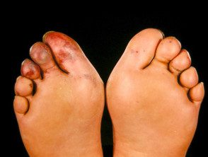
Peripheral gangrene due to ischemia
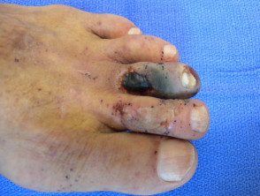
Dry gangrene due to ischemia
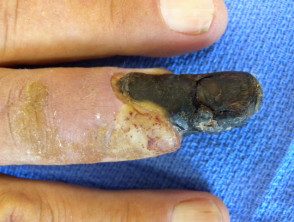
Dry gangrene due to ischemia
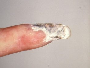
Dry gangrene due to ischemia
What are the first signs of ischemia?
Proximal severe acute Ischemia presents as a pale, paralyzed, pulseless limb. This is a surgical emergency because it can progress to extensive gangrene if the obstruction is not cleared quickly.
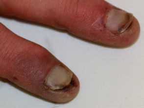
Distal peripheral vascular the blockage appears as blue, gray, or purple patches, blisters, or ulcers at the end of the digits. See blue tip syndrome.
Ischemia at other sites can also result in localization necrosis of the skin and deeper tissues.
Peripheral ischemia
What Causes Ischemia?
Ischemia is due to an acute or chronic interruption in blood supply and is often due to a combination of both.
Acute ischemia can be due to:
- Embolism
- Acute arterial thrombosis
- Vascular trauma
- Calciphylaxis
- Extreme cold injury (frostbite)
- Infection-related disseminated intravascular coagulopathy or purple fulminants
Emboli
Emboli are particles that flow through the bloodstream until they lodge in an arterial junction or narrowing. Emboli can be composed of:
- Cholesterol Dislodged from the heart (80%) or a large artery (aorta, iliac vessel)
- Tumor
- Infectious particles
- Fat shed during an operation
- Strange objects.
Large emboli lodge in the bifurcation of the femoral artery (43%), the iliac arteries (18%), the aorta (15%), and the popliteal arteries (15%). The little ones are deposited on the fingers and toes, palms and soles of the feet.
Vascular obstruction may also be due to hyperviscosity syndromes or abnormal circulating proteins such as cryoglobulins or macroglobulins.
Thrombosis
Thrombosis is a blood clot that arises within a blood vessel. Predisposing factors include:
- Septicemia
- Hypotension (low blood pressure)
- Low cardiac output
- An aneurysm (a bulge in the wall of a weakened artery)
- Aortic dissection (a division in the wall of the aorta)
- Bypass graft (a surgical procedure)
- Atherosclerotic narrowing of the artery.
Whatever the primary Cause of ischemia, thrombosis due to slow blood flow can make it worse.
Chronic ischemia can be due to:
- Peripheral arterial disease
- Venous insufficiency.
Peripheral arterial disease
Chronic peripheral arterial disease is most often due to atherosclerosis. This is a buildup of cholesterol, fibrin and other proteins within the arterial wall, causing the vessel to narrow and eventually become completely blocked. This process is also called obliterative arteriosclerosis.
Risk factors for peripheral arterial disease include:
- Hypertension (hypertension)
- Hyperlipidemia (high fats in the blood)
- Diabetes
- Cardiovascular disease including myocardial infarction, atrial fibrillation, valve disease
- Cerebrovascular disease including stroke, transient ischemic attack
- Of smoking
- Autoimmune diseases: vasculitis, arthritiscoagulopathy
- Malignancy (Cancer)
Peripheral arterial disease can also be caused by:
- Connective tissue disease, including systemic sclerosis
-
Buerger's disease (obliterative thromboangitis).
Venous insufficiency
Veins may fail to remove blood from tissues due to valve dysfunction (varicose veins) and obstruction by deep vein thrombosis (DVT) or thrombophlebitis. High pressure within the veins (venous hypertension) results in the movement of proteins and fibrin into the Soft fabric. This leads to fibrosis, dysfunctional capillary fat formation and necrosis (lipodermatosclerosis). When severe, these processes can cause ischemia and gangrene.
Clinical characteristics of vascular obstruction.
Signs of peripheral vascular disease depend on which tissues are ischemic and their severity. A patient may present with:
- Claudication (intermittent pain in the leg)
- Pain at rest
- Shiny, thin, dry, pale and cold skin.
- Fragile nail
- The most advanced disease can be revealed by livedo reticularis (network appearance)
- Bad healing and ulceration (most often leg ulcers)
- Gangrene: an area of gray or black necrotic (dead) tissue.
Investigations in a patient with ischemia or dry gangrene.
When a patient has peripheral ischemia or gangrene of unknown cause, a complete physical examination is performed to evaluate the vascular system, including the heart and peripheral pulses. An electrocardiogram (ECG) assesses heart function. Blood pressure is measured with Doppler ultrasound of both lower extremities and both upper extremities to calculate the ankle brachial index (ABI).
- ABI 1–1.4 is normal when pressure in the lower extremities is equal to or greater than in the upper extremities.
- ABI 0.5–0.8 indicates mild to moderate arterial disease.
- ABI> 0.5 indicates severe arterial disease.
Images to evaluate peripheral vascular disease may include:
- Vascular ultrasound: to locate the site or sites of obstruction in arteries and veins.
- Angiography - a dye scan to identify the type, location and extent of vascular obstruction in the affected limb.
- Chest x-ray
- Magnetic resonance (Magnetic resonance) - which can replace traditional angiography.
- Computed tomography (Connecticut) - this may reveal calcification and with contrast (dye) it can reveal vascular obstruction.
- Echocardiogram - ultrasound of the heart valves to identify a source of emboli.
Blood tests assess blood count, kidney function, electrolytes, lipid profile, coagulation state and inflammatory markers such as D-dimer and C-reactive protein.
What is the treatment for ischemic gangrene?
Gangrene treatment varies by location and cause, but focuses on radical surgery. debridement +/- amputation. Surgical procedures can also include:
- Elimination of plunger or thrombus
- Balloon or stent catheterization
- Arterial or venous bypass surgery
- Hyperbaric oxygen treatment.
Medical treatment may include:
-
Antiplatelet agents, including aspirin, clopidogrel, cilostazol, and pentoxifylline.
- Lipid-lowering agents such as statins
- Treatment of underlying conditions such as diabetes.
- Cessation of medications that cause vasoconstriction
- Give up smoking
- Avoid exposing the affected area to cold.
Which is the forecast for gangrene?
The prognosis of ischemic gangrene depends on the extent of the disease, the underlying cause, and the timing of appropriate treatment.
There will be scarring and there may be some reduction in function, especially if significant debridement or amputation has been necessary. Reappearance of ischemia is probably due to medical comorbidity, especially diabetes.
