
Introduction
Glomangiomas (also called glomuvenous malformation) differ clinically from glomus tumors in that they occur in childhood and adolescence, generally asymptomaticdoes not have a predilection For him subungic region, and are often multifocal. They can vary in color from pink to blue and often get darker with age; They may be license plate-like or nodular. Multiple glomangiomas are rare and comprise approximately 10 percent of all glomus tumors.
Histology glomangioma
In glomangioma, the histopathology dilated sample venous channels that resemble venous malformations (Figures 1, 2). Unlike venous malformations, they demonstrate one to several rows of cells from surrounding cuboidal blood cells (Figures 3,4).
Glomangioma pathology
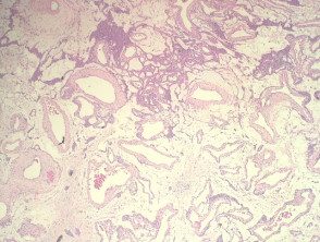
Figure 1
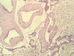
Figure 2
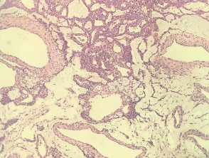
figure 3
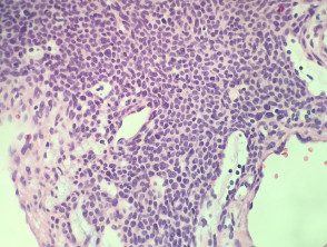
Figure 4
Glomal cells stain positively for vimentin and smooth muscle α actin, but are negative for desmin, von Willibrand factor, and S-100.
Differential diagnosis for glomangioma
Other diagnoses to consider include:
- Vascular malformation - these do not have the perivascular Glomal cell accumulations are seen in the glomangioma.
- Myopericytoma, angioleiomyoma, myofibroma - they all have varying degrees of perivascular muscle cells, but these cells lack the classic cuboidal glomus cells. These tumors generally show a degree of desmin positivity in the perivascular cells that are not seen in the glomangioma.
