What is childish? proliferative hemangioma?
Infantile hemangioma (American spelling “hemangioma”) describes a benign condition (non-cancerous) that affects cutaneous blood vessels. It is known as a proliferative hemangioma because it is due to proliferating endothelial cells (the cells that line blood vessels).
Infant proliferative hemangiomas usually develop shortly after birth. They are different from vascular malformations, which are generally present at birth and are less common.
More than 80% of infantile proliferation hemangiomas occur in the head and neck area. They grow to 80% of maximum size in the first three months and most stop growing within 5 months. However, they can continue to grow for up to 18 months.
After that, they submit regression or involution This can take 3 to 10 years. Most flat infantile proliferative hemangiomas eventually involve and disappear without treatment. However, the regression of bulky hemangiomas tends to be incomplete and may leave an irregular discharge. atrophic scar or anetoderma (a dented scar) in at least 50% of cases.
Types of infantile proliferative hemangioma
Infantile proliferative hemangiomas are classified as superficial, deep, or mixed lesions. They may be located or segmental (which implies a larger neuroectodermal unit).
- Superficial infantile hemangioma is also called capillary capillary hemangioma nevus, strawberry hemangioma, strawberry nevus and simple hemangioma. The blood vessels in the upper layers of the skin are dilated.
- Deep infantile hemangiomas are also called cavernous hemangiomas and are established deeper in the dermis and subcutis. They appear as a soft to firm bluish swelling.
- Both types of hemangiomas can occur together in mixed angiomatous naevi when a strawberry nevus covers a bluish swelling.
Segmental proliferative hemangiomas are more serious than localized hemangiomas.
- They occur at a younger age and grow up to ten times larger
- Consequently, they are more unsightly.
- Other congenital abnormalities may be associated with facial segmental hemangiomas (PHACE syndrome) These include later fossa abnormalities, hemangiomas, arterial abnormalities, cardiac abnormalities and eye abnormalities.
- Likewise, segmental infantile hemangiomas involving perineum may be associated with congenital pelvic abnormalities, PELVIS syndrome (perineal hemangioma with any of the following: external genital malformations, lipomyelomeningocele, vesicorenal abnormalities, imperforate anus, or skin tag)
Capillary hemangiomas
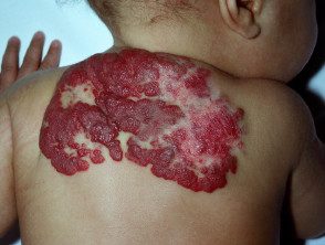
Infantile proliferative hemangioma
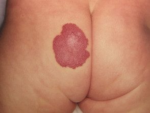
Hemangioma
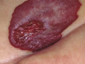
Hemangioma
Cavernous hemangiomas (mixed type)
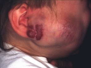
Hemangioma
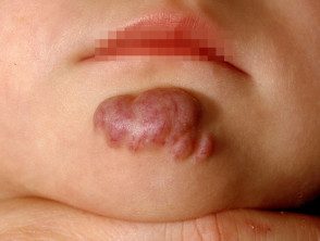
Hemangioma
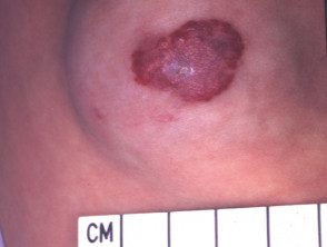
Hemangioma
Other hemangiomas
The hemangiomas described below are very rare conditions.
| Kind | Characteristics |
|---|---|
| Warty hemangioma |
|
| Eruptive neonatal hemangiomatosis |
|
| Mutilating ulcerative hemangiomatosis |
|
| Acquired multiple hemangiomatosis |
|
Kasabach-Merritt syndrome is also known as hemangioma.thrombocytopenia syndrome. It is a rare complication of a rapidly growing vascular lesion, but is no longer believed to arise from a common infantile proliferative hemangioma.
What Babies Have Proliferative Hemangiomas?
Ten percent of babies develop one or more hemangiomas. Localized hemangiomas are more common if the baby is low birth weight, for example under the following circumstances.
- Female
- White skin
- Premature
- Multiple births (twins)
- Advanced maternal age
- Family history of infantile proliferative hemangioma
Hypoxia (inadequate oxygen for the skin) is now considered the probable reason for the proliferation of blood vessels Endothelial progenitor cells (EPCs) circulate in a fetus and cause new blood vessels to form in response to hypoxia. Typically, CLDs are gone when a baby is born, but can still be present in low-birth-weight or premature babies. As the EPCs disappear later in life, the hemangioma may regress.
Segmental hemangiomas are believed to arise early in gestation (6-8 weeks) as a developmental error.
Investigations in infants with infantile proliferative hemangioma
Infantile hemangiomas are generally diagnosed clinically and investigations are not needed for most superficial lesions.
Deep infantile hemangiomas or segmental hemangiomas are routinely investigated with ultrasound exploration. An ultrasound is also often done when there is uncertainty about the diagnosis or if underlying tissues are affected. Characteristically, a hemangioma has a firm lobular structure with vessels separating the lobules.
It may also be necessary to perform Magnetic resonance imaging (Magnetic resonance) or angiography to help plan treatment. Children with complex injuries are best evaluated by a panel of experts, including the pediatrician, dermatologistradiologist ophthalmologist, vascular and plastic surgeons.
Hemangiomas that arise on the lower spine are sometimes a marker of hidden spinal dysraphism (spina bifida), when spinal imaging may be appropriate.
When is treatment for infantile proliferative hemangioma necessary?
Because infantile hemangiomas are likely to improve or come back completely over time, there is no need for specific treatment in most cases. Treatment should be considered in the following circumstances.
- Very large and unsightly lesions.
- Ulcerating hemangiomas (up to 5–25% of lesions)
- Injuries that affect vision, hearing, breathing, or eating.
- If not resolved by school age
- Hemangiomas that have a steep or stepped edge, a coarse pebble surface, or superficial and deep components combined.
The baby is best evaluated early during the rapid growth phase at the age of 3 to 5 months. If the injury obstructs vision, it can prevent the development of normal vision.
What is the treatment for infantile proliferative hemangioma?
Propranolol is the treatment of choice for problematic hemangiomas. Current beta-blockers such as timolol, available as eye drops or 0.5% gelforming solution, can be used off-label for small superficial hemangiomas.
Other possible treatments include:
- External compression therapy (bandage)
- Ultra Powerful Topical Steroids
-
Topical antiseptics. Eosin, which also has antiangiogenic properties, has been reported to be beneficial.
-
High-dose oral corticosteroids, during the proliferative stage of segmental disease (mostly replaced by propranolol)
- Sometimes intralesional steroid injections have been used for small hemangiomas.
-
Vascular To be therapy at the age of 3 to 4 years, when the lesions are stable
- Interferon alpha may be helpful, but is rarely recommended as it has been associated with the development of cerebral paralysis in some infants.
- Vincristine was reported to be effective in the past, but is rarely used today
-
Imiquimod has been reported to accelerate resolution in some cases.
