
What is oral (irritated)? fibroma?
An oral fibroid is common benign scarreaction similar to persistent and persistent irritation in the mouth. Also known as traumatic fibroma, focal intraoral fibrous hyperplasiafibrous nodule or oral polyp.
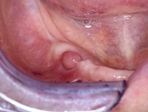
Oral fibroma
Oral fibroma
Who gets an oral fibroid?
An oral fibroid is most often seen in older adults, but it can occur at any age. It affects 1–2% in adults.
It is generally due to chronic irritation such as:
- biting your cheeks or lips
- rub off a rough tooth
- dentures or other dental prostheses.
What are the clinical features of an oral fibroma?
An oral fibroma presents as a smooth firm papule in the mouth. It is usually the same color as the rest of the lining of the mouth, but it is sometimes paler or, if it has bled, it may appear dark in color. The surface may be ulcerated due to traumaor get rough and scaly. It is usually dome shaped, but may have a short stem like a polyp (pedunculated) If it has developed under dentures, it may be flat in the shape of a leaf.
The most common location for an oral fibroid is on the inside of the cheek, where the upper and lower teeth meet. Other common sites include the sides of the tongue, the gums, and the inside of the lower lip.
Aside from feel and appearance, oral fibroids It does not cause any symptoms. Oral fibroids develop over weeks or months to reach a maximum size, usually about 1 cm in diameter, but can sometimes be larger.
An oral fibroid is usually a loner. injury. When there are many lesions, associated diagnoses should be considered, including tuberous sclerosisCowden syndrome, family fibromatosis and fibrotic papillary hyperplasia of the palate.
Oral fibroids do not turn into oral Cancer.
In addition to fibroid irritation, there are other well-recognized types of oral fibroma:
- Oral elastofibroma
- Epulis fissuratum
- Giant cell fibroma
- Myofibroma and myofibromatosis
- Peripheral ossifying fibroma
- Peripheral odontogenic fibroma
- Retrocuspid papilla
- Sclerotic fibroma.
How is oral fibroma diagnosed?
The diagnosis of oral fibroma will be suspected clinically when there is the usual history and the results of the examination. A biopsy it can be taken to exclude other conditions or to eliminate injury. Histology shows typical dense fibrous tissue with relatively few cells. The overlying epithelium it may be ulcerated, thin, or thickened.
What is the treatment of oral fibroma?
When treatment is required, the only option is surgical excision fibroma with narrow margins. It may come back after surgery if the source of irritation continues. Therefore, it is also important to control the source of the irritation. Oral fibroids do not go away without treatment.
