
What is it youth idiopathic arthritis?
Juvenile idiopathic arthritis (JIA) covers everything chronic childhood arthropathies beginning before age 16 with persistent arthritis for at least 6 weeks.
JIA It is recognized for having seven main subtypes, some of which exhibit the clinical and pathological characteristics of others. autoimmune Disorders These subtypes include:
- Systemic JIA (also known as juvenile onset disease)
- Oligoarthritis
- Positive rheumatic factor (RF) polyarthritis
- RF negative polyarthritis
- Psoriasic arthritis
- Enthesitisrelated to arthritis
- Undifferentiated arthritis [1,2].
Idiopathic juvenile rheumatoid arthritis
Arthritis
Exanthem
Exanthem
Who gets Juvenile Idiopathic Arthritis?
JIA can occur in anyone from infants to teens and has an average age of onset of 6 years. Most types of JIA affect boys and girls equally, although systemic-onset JIA (juvenile-onset disease) is predominantly seen in girls [3].
What causes juvenile idiopathic arthritis?
The exact cause of JIA is unclear, but a pronounced immune response in a perpetual cycle of adaptive and innate immunity is believed to contribute to Pathogenesis from JIA. Aberrant activation of the innate immune system leads to deregulated production of proinflammatory cytokines. Tumor necrosis alpha factor (TNF-α), interleukin (IL) -1 and IL-6 play a critical role in incitement. The effects of these cytokines provide explanations for the various clinical features observed in systemic JIA. These effects include:
- Granulopoiesis of the bone marrow (proliferation of granulocytes)
- Increased osteoclastic activity (cells that absorb bone).
- Hepatocyte stimulus
- Activation of thermoregulatory functions (body temperature) [3,4].
Increased recruitment of inflammatory cells in the joint synovial the membrane was demonstrated in several studies by a significant elevation of proinflammatory cytokines in the synovial fluid of affected children. Proinflammatory cytokines in the synovial lead to increased production and accumulation of synovial fluid and thickening of the synovial lining. This chronic synovium inflammation it is apparent and common to all JIA subtypes. Furthermore, RANKL (receiver activator nuclear kappa factor-Β ligand) cytokine, which is associated with bone resorption (decomposition) and cartilage damage, was reported to be present in large amounts in the synovial membrane of children with JIA.
Some studies have consistently supported associations between enthesitis-related JIA and human White blood cell antigen (HLA) polymorphisms; There appears to be an increase in associations, especially with early onset disease. A monogenic form of systemic JIA with an underlying mutation in a regulatory protein involved in macrophage metabolism has been described [2,3].
Environmental factors can contribute to the development of JIA. Some studies have suggested associations with exposure to antibiotics, a bacterial infection in immunosuppressed individuals and maternal smoking during pregnancy [2,4].
What are the clinical features of juvenile idiopathic arthritis?
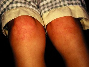
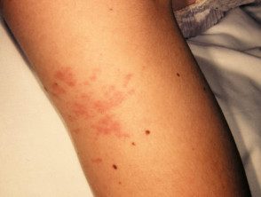
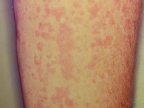
Cutaneous Participation is one of the key extra-articular factors in systemic JIA. the exanthema or eruption is presented as discreet, Salmon pink maculesor edematous papules They are usually transitory and coincide with fevers. These tend to occur on the trunk, armpitsand proximal extremities, but not commonly seen on palms, soles, or face. Persistent plates, periorbital edemaor erythema they are less common but can be observed.
Although eruptions at JIA rarely pruritus, can be caused by skin lesions or scratching (Koebner phenomenon), in which case the injuries exhibit a linear appearance.
Children with JIA often have a limp and stiffness, without any known injury or previous infection. Common non-cutaneous manifestations include:
- Joint pain and inflammation
- Daily high peak fever (daily model)
- Myalgia
- Limited movement
- Enthesitis
- Sore throat
- Lymphadenopathy
- Hepatosplenomegaly [1–3].
What are the complications of juvenile idiopathic arthritis?
Musculoskeletal complications secondary to continuous inflammation and poor disease may occur control, including:
- A leg length discrepancy
- Joint erosion
- Sacroiliac joint and ankylosis of the spine (stiffness due to bone fusion).
Sometimes JIA can lead to more serious complications with devastating results. These may include:
- Uveitis - this can lead to loss of vision; screening and close monitoring by a pediatric ophthalmologist is guaranteed for severe active uveitis
- Macrophage activation syndrome (MORE): This can be deadly; characterized by fever, cytopenias, liver dysfunction, coagulopathy, purple, hypofibrinogenesis (a disorder of blood clotting factor), hypertriglyceridemia (high levels of triglycerides in the blood), and a very high level of ferritin [2,3].
How is juvenile idiopathic arthritis diagnosed?
The diagnosis of systemic JIA is made clinically after the exclusion of other entities, such as malignant tumors and infections that can also present with high fever and arthralgia. According to the criteria of the International League of Rheumatology Associations (ILAR) for systemic JIA, arthritis must have persisted for at least 6 weeks and fever for 2 weeks, accompanied by one or more of the following:
- Transient erythematous eruption
- Generalized lymphadenopathy
- Hepatosplenomegaly
- Serositis (inflammation of a serous membrane) [5].
When JIA is suspected, pediatric rheumatologists should be immediately involved to initiate appropriate investigations and minimize any delay in treatment. Additional research, including laboratory tests and imaging, is helpful in confirming the diagnosis or classifying arthritis into a specific subtype. Test results can also be helpful in guiding treatment and monitoring the course of the disease.
The most relevant laboratory tests for JIA are:
- A complete blood count anemia, thrombocytosis, and leukocytosis are often present in systemic JIA, while not typically seen in oligoarticular JIA
- Erythrocytes sediment speedESR): This is significantly elevated in systemic JIA, but may be normal or mild to moderately elevated in other subtypes.
- C-reactive protein (CRP) - this is raised to varying degrees in different JIA subtypes
- Antinuclear antibody (ANA): This is often negative in systemic JIA, but may be positive in oligoarticular and polyarticular forms of JIA
- RF and anticyclic citrulline peptide (anti-PCC); These are useful in identifying RF-positive polyarthritis.
Pathology not required for a definitive diagnosis of JIA, but the rash in systemic JIA characteristically exhibits a perivascular and interstitial neutrophils-dominant infiltrate; this can also be called neutrophilic urticarial skin disease, a common pattern in autoinflammatory processes [3].
The images may provide information about the joints involved if the clinical examination findings are equivocal. A ultrasound exploration can assess synovitis in the joints and magnetic resonance image can be used to rule out other conditions like pigmented Villonodular synovitis (inflammation of the joint lining) [2].
Which is the differential diagnosis for juvenile idiopathic arthritis?
Several conditions can present similarly to systemic-onset JIA, including having a transient rash, fever, and arthritis. These include:
- Rheumatic fever
- Urticaria vasculitis
- Serum disease reaction
- Juvenile dermatomyositis
- Systemic lupus erythematosus
-
Kawasaki disease [3].
What is the treatment for juvenile idiopathic arthritis?
Treatment of systemic JIA is aimed at symptom management, improvement or management of joint function and minimization of complications. Nonsteroidal anti-inflammatory drugs (NSAID) are usually the first-line agents for mild joint or extra-joint presentations to control pain. Other pharmacological agents include:
-
Oral corticosteroids for moderate to severe disease.
-
Intravenous corticosteroids: may be necessary in patients with outbreaks or serious complications, such as MAS.
- Disease modifying antirheumatic drugs (DMARDs), such as methotrexate, cyclosporine, and leflunomide.
Medications that block inflammatory cytokines, such as TNF-α, IL-6, and IL-1, have been shown effectiveness, especially in long-standing JIA. These include:
-
Anakinra: an IL-1 receptor antagonist
- Canakinumab - a humanized monoclonal antibody directed against IL-1
- Tocilizumab: This humanoid monoclonal antibody blocks IL-6 [2–4].
What is the result for juvenile idiopathic arthritis?
The course of systemic JIA disease is variable. Reports have indicated that between 40 and 60% of patients reach the remission arthralgia Systemic manifestations may remain active and persist after years. A better forecast it is associated with early treatment of active disease [2,3].
