
What is a leiomyoma?
A leiomyoma is a benign tumor composed of smooth muscle. It is capable of arising wherever smooth muscle is present. One form of leiomyoma arises from uterine smooth muscle, and is also known as uterine fibroids.
Cutaneous Leiomyomas can be classified into three types:
- Piloleiomyoma
- Angioleiomyoma
- Genital leiomyoma
Each type arises from smooth muscle in specific tissues or organs and has distinct clinical or clinical characteristics. histological features.
Piloleiomyoma
- It originates from the retractor pili muscle of the pilosebaceous unit.
- Single or multiple injuries.
- Multiple lesions can occur sporadically or are inherited together with uterine fibroids in a autosomal dominant pattern as part of hereditary leiomyomatosis and renal cell Cancer syndrome or as part of Reed's syndrome (multiple cutaneous and uterine leiomyomatosis)
Angioleiomyoma
- Originate in vascular smooth wall muscle tissue
- Typically occurs as a loner injury
Genital leiomyoma
- It originates from the dartos muscle in the scrotum or labia majora, or from the erectile muscle in the nipples.
- Typically occurs as a solitary injury
- Less common cutaneous leiomyoma
Who gets leiomyomas?
Uterine leiomyomas represent 95% of all reported leiomyomas. Cutaneous leiomyomas account for 75% of all extrauterine leiomyomas.
- Incidence it is largely unrelated to race.
- Most solitary cutaneous leiomyomas occur in adulthood. Multiple piloleiomyomas usually occur between the ages of 10 and 30.
- Excluding autosomal dominant syndromes, the incidence of piloleiomyoma is equal in men and women. Angioleiomyomas are generally more common in women than men (2: 1) although cavernous and venous The subtypes are more common in men.
What are the clinical features of leiomyomas?
Leiomyomas are often painful.
- Pain can be spontaneous or triggered by physical and / or emotional stimuli, including temperature or cold pressure. Menstruation or pregnancy can also act as triggers
Piloleiomyomas
- Likely to present with associated pain
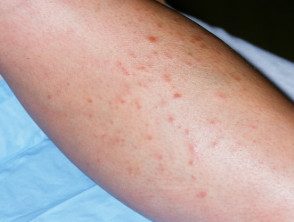
- Typically cuddly, mobile hyperpigmented or reddish brown nodules (≤ 2 cm in diameter) with a smooth surface and firm consistency
- Solitary lesions typically on the lower extremities, while multiple piloleiomyomas appear anywhere in a variety of distribution patterns
Angioleiomyomas
- 60% reported to be painful
- Tenderness is less common than in piloleiomyomas.
- All right circumscribed, skin color, solitary nodules ≤ 4 cm in diameter, usually found on the lower legs. Uncommon on the head, trunk, hands or mouth.
Genital leiomyomas
- Typically not tender and painless
- Firm, solitary, skin-colored, mobile. nodule about him vulva, scrotum or nipple. The size is more variable than other types; Vulvar and scrotal lesions are typically larger.
Leiomyoma
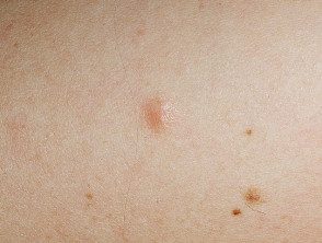
Leiomyomas
How are cutaneous leiomyomas generally diagnosed?
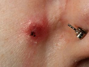
Cutaneous leiomyomas are usually diagnosed by the skin. biopsy. Each type of leiomyoma has unique characteristics. histology.
How are leiomyomas treated?
Surgical excision It is the definitive treatment for individual injuries. Multiple cutaneous leiomyomas have a high rate of reappearance (~ 50%) in weeks or years, especially if it is part of HLRCC or Reed syndrome.
Medical treatment is not curative, but nifedipine, phenoxybenzamine, and gabapentin can relieve pain.
