
What is it cancel lichenoid dermatitis Of the Youth?
Lichenoid annulare dermatitis of youth is a rare skin condition that was first described in 2003 [1]. It is characterized by sharply demarcated cancel erythematous macules and papules with central hypopigmentation [2].
Who gets lichenoid annular dermatitis in youth?
Lichenoid annulare dermatitis of youth was first described in healthy young individuals from the Mediterranean. [1]. The average age of patients affected by this skin condition is 10 years. [2]. Cases have been reported in adults and older people from central Europe, the US, and Japan [3,4].
What causes lichenoid annulare dermatitis of youth?
The exact cause of lichenoid annulare dermatitis in youth has not yet been discovered. It is believed to share pathogen factors with other lichenoids skin disease, such as lichen planus, graft versus host lichenoid disease and drug eruption, which involve a T cell-mediated and subsequent response apoptosis of basal keratinocytes [2]. The reasons why challenge the ridges are directed in lichenoid annulare dermatitis are currently unknown.
What are the clinical features of lichenoid annulare dermatitis in youth?
Lichenoid annulare dermatitis usually presents as asymptomatic Red-brown round, oval, or annular macules or patches. These begin as erythematous macules with a raised border and develop central hypopigmentation as they gradually enlarge (Figure 1) [2]. Macules or patches do not itch.
Lesions are found on the patient's trunk, groin and, less frequently, on the armpits [2]. The individual is fine otherwise, without systemic symptoms seen
Figure 1. Lichenoid annular dermatitis of youth.
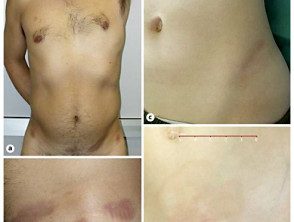
Lichenoid annular dermatitis
Credit: Prof Paolo Gisondi
How is lichenoid annulare dermatitis diagnosed in young people?
Lichenoid annulare dermatitis of youth is diagnosed in histopathology, with the presence of specific distinctive features. These features include:
- A interface dermatitis only affects the tips of the ridge crests (Figure 2) [2]
- A loss of keratinocytes, which results in a dulling of the rete ridges, giving a square appearance (Figure 2) [2].
Figure 2. Histopathological characteristics of lichenoid annulare dermatitis of youth.
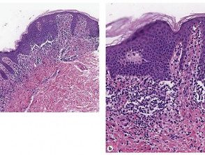
Histopathology of lichenoid annular dermatitis
Credit: Prof Paolo Gisondi
Which is the differential diagnosis for lichenoid annulare dermatitis of youth?
The lichenoid annulare dermatitis of youth can be confused clinically with morpho, mycosis fungoides and annulare erythema.
-
The morpho is differentiated For this histopathological findings of a atrophic epidermisthickened hyaline collagenor loss of sweat glands and hair follicles.
- Hypopigmented mycosis fungoides differentiates histopathologically for him infiltration of evil one T cells in the epidermis and by extensive spongiosis.
-
Erythema annulare usually resolves on its own and often scaly. Histopathological findings show a inflammatory cell infiltrate Surrounding surface vessels, known as the 'coat sleeve anomaly'.
What is the treatment for lichenoid annulare dermatitis of youth?
Transient referrals can be achieved in lichenoid annular dermatitis of youth with treatment with current steroids, systemic steroids, topical tacrolimus, or narrowband ultraviolet B phototherapy [2].
Lesions may reappear once treatment has been stopped. [2]. Spontaneous recovery without treatment can also occur, but this is rare.
What is the outcome for lichenoid annulare dermatitis of youth?
Lichenoid annulare dermatitis of youth progresses slowly, with no serious complications reported to date. The duration of lichenoid annulare dermatitis of youth varies. Of the 21 cases described between 2005 and 2013, most of the lesions were successfully treated with topical corticosteroids, but these recurred once treatment was discontinued. [2]. There have been some cases that demonstrated complete remission after treatment cessation, and a case that showed spontaneous regression without any treatment [2].
