
What is a lymphatic malformation?
A lymphatic malformation is a type of vascular nevus or birthmark due to malformed and dilated lymphatic vessels.
What is the cause of lymphatic malformation?
The cause of malformed dilated lymphatic vessels is not fully understood. The embryonic lymphatic sacs have not connected properly with the lymphatic system for some reason. They are not cancerous
Who gets lymphatic malformation?
No particular race or sex You are more at risk.
What are the clinical features of lymphatic malformation?
Lymphatic malformations are always present at birth, although they can be missed; they tend to become more obvious during infancy and childhood. They can range in size from a small point to occasionally involving an entire limb. Although lymph is a clear fluid, lymphatic malformations often appear red or blue due to the involvement of adjacent blood vessels.
Classification of lymphatic malformation.
There are three main types of lymphatic malformation:
- Cystic hygroma
- Cavernous lymphangioma
- Lymphangioma circumscriptum
Cystic hygroma
Cystic hygroma (also called “cystic lymphangioma” and “cystic lymphangioma”) is a “macrocytic” lymphatic malformation and is made up of large fluid-filled spaces. It appears as a colored, red or bluish skin, somewhat transparent, swelling under the skin. Cystic hygroma most often affects the neck, armpits, or groin. Very large lesions can affect the chin and face, and occasionally interfere with eating or breathing. They can become infected, which can be life-threatening if the neck is involved.
Cystic hygroma is more common in some congenital disorders including turner syndrome, hydrops fetalis, Down syndrome and fetal alcohol syndrome. Therefore, it may be advisable for babies with this type of lymphatic malformation to have chromosomal Analysis performed.
A ultrasound The scan is often done to find out the extent of the abnormality. Characteristically, firm rounded spaces are detected. A cystic hygroma is distinguished from venous malformation and hemangioma by the appearance and lack of fluid flow.
In more complicated cases it may be necessary to perform Magnetic resonance imaging (Magnetic resonance) or angiography to help plan treatment.
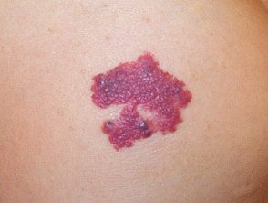
Cavernous lymphangioma
Cavernous lymphangioma can affect any site on the body, including the tongue. It appears as a red, red, or bluish swollen skin under the skin. Sometimes it has a period of rapid growth in early childhood. In rare cases, it can ulcerate. Differs from other vascular naevi by the presence of clear fluid within the lumps and findings on ultrasound.
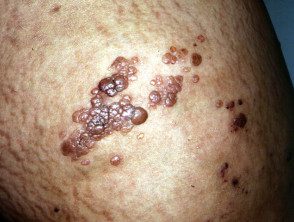
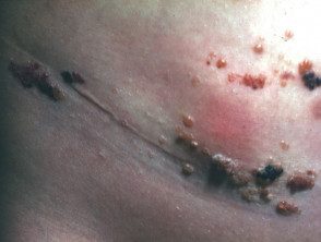
Lymphangioma circumscriptum
Lymphangioma circumscriptum is a "microcytic" lymphatic malformation (see Lymphangioma circumscriptum histology) Appears as a group of small firm blisters filled with lymphatic fluid, resembling hatched frogs. These vary in color from light to pink, dark red, brown or black and can turn warty, especially when it affects a palm or sole. Lymphangioma circumscriptum is most commonly found on the shoulders, neck, underarm area, extremities, and in the mouth, especially on the tongue. The patches may become more prominent at puberty and may ooze or bleed. They occasionally become infected.
Lymphangioma circumscriptum
Lymphangioma
Lymphangioma
Lymphangioma
What treatments are available for lymphatic malformation?
Cystic hygroma and cavernous lymphangiomas are usually removed surgically. Unfortunately, deeper lesions are quite difficult to remove completely, and sometimes they grow back. Oral sirolimus (which inhibits angiogenesis) and sildenafil (which causes dilation of blood vessels) are being investigated as possible medical treatments for severe lymphatic malformations, but may have significant adverse effects [2,3].
Lymphangioma circumscriptum is benign (non-cancerous) and generally does not require treatment. Antibiotics may be given if secondary infection happens (rare). If necessary, it can be removed surgically, by dermabrasion, or by To be. They have a tendency to reappear after treatment.
