
What is a nevus?
A nevus (American spelling nevus, nevi) is a circumscribed and stable malformation of a skin component. Naevi (American spelling Nevi) are often present at birth, when they are often called brown birthmarks. Naevi composed of melanocytes (the pigment cells that produce melanin) They are called melanocytic naevi or pigmented naevi.
What are Ota, Ito and Hori naevi?
Nevi of Ota, Nevi of Ito, and Nevi of Hori are special melanocytic nevi that have a slate brown or blue / gray coloration. They are forms of dermal melanocytosis in which nevus cells lie deep within the dermis.
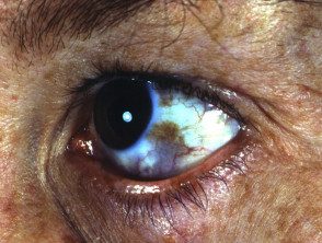
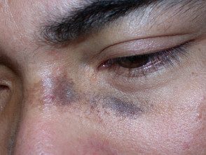
- Ota's Naevus is on the forehead and face around the eye area. Hyperpigmentation parts of the eye can occur (sclerotic, corneairis, retina)
- Naevus of Hori is similar to Naevus of Ota but affects both sides of the face
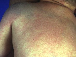
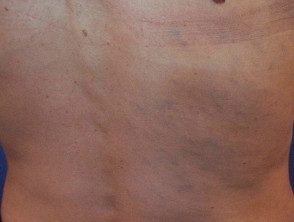
- Ito's naevus is in the shoulder and upper arm area (shoulder girdle).
Dermal melanocytosis can also occur in other parts of the body, including inside the mouth.
Naevus of Ota
Naevus of Ota

Naevus of Ota
Naevus of Ota
Naevus by Ito
Naevus by Ito
Naevus by Ito
How does dermal melanocytosis arise and who is at risk?
It is not known why dermal melanocytosis occurs. Specific mutations they have been detected within dermal melanocytes, most frequently GNAQ or GNA11. Researchers have suggested that hormones play a role in its development. The role of Ultraviolet radiation it is believed to be small, as it does not reach deep dermal melanocytes.
Ota's naevus is much more common than Ito's neevus. These nevi are present at birth in 50% of cases, but can appear during adolescence or adult life. Naevus of Hori is not present at birth and is therefore a form of acquired melanocytosis.
Naevi de Ota and Ito are most commonly found in Asian populations; 0.2-0.6% of the Japanese have a nevus of Ota. They appear more often in women. Both forms of nevi are rare in Caucasians.
What are the signs, symptoms, and complications of dermal melanocytosis?
In all forms of dermal melanocytosis:
- Color may vary to include brown-purple, purple-blue, or blue-green shades
- Naevi present in childhood can slowly grow and darken until they reach adulthood.
- The color or perceived color of nevi may change according to personal and environmental conditions, eg. fatigue, menstruation, hot weather
- If it affects the eye, melanocytosis rarely causes glaucoma
- Evil one melanoma it very rarely develops within dermal melanocytosis, and has generally been reported in Caucasians. Ocular Melanoma of the choroid, brain, orbit, iris, ciliary body, and optic nerve has rarely been reported in association with a nevus of Ota.
How is the diagnosis of dermal melanocytosis made?
The diagnosis of dermal melanocytosis is usually made by looking at the typical discoloration of the skin. It is classified according to the affected site.
Some patients can undergo the skin. biopsy, which confirms the presence of melanocytes in the dermis.
Other skin conditions that result in bluish or gray skin can be considered. These include:
- Blue nevus
-
Drug induced pigmentation, most often due to minocycline, a tetracycline antibiotic
- Melasma
- Post-inflammatory pigmentation
-
Pigmented and ashy lichen planus skin disease
What treatments are available for melanocytosis?
Treatment for a melanocytosis may include cosmetic camouflage to cover the disfiguring marks, and To be treatment (generally using 1064nm Q changed Nd: YAG, Alexandrite or QS ruby laser) or intense pulsed light (IPL).
-
The picosecond laser may be the most effective device.
- Laser and light devices work by destroying dermal melanocytes.
- Multiple treatments are necessary, often with a combination of devices.
- Laser treatment is more effective in treating melanocytosis in fair-skinned individuals compared to those with dark skin.
- Reappearance is common after laser lightening, sometimes it results in a darker shade than before treatment.
If the eye is affected, regular eye exams should be arranged to detect glaucoma. Any change in a nevus should be evaluated by a dermatologist.
