
What is a blue nevus?
A blue nevus is a common type of melanocytic nevus in which the pigment is located in the background dermis. Also known as a blue neuronaevus and a dermal melanocytoma
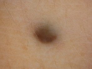
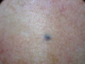
What are the clinical characteristics of a blue nevus?
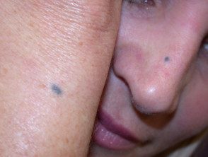
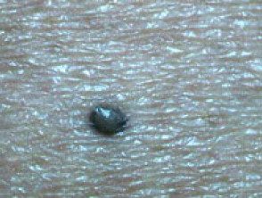
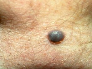
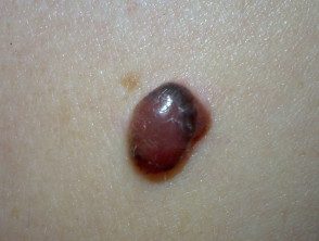
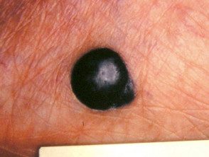
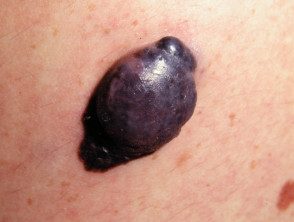
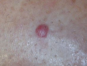
A blue nevus is a well.circumscribed round or oval taint, papule or nodule of a uniform steel blue color. The edge gradually fades into the surrounding skin.
- The common blue nevus is usually 0.5–1 cm in diameter. the cellular blue nevus is over nodular and is at least 1 cm in diameter.
- The color of blue naevi It can also vary, generally consisting of blue to gray tones, but sometimes they are brown or yellowish.
- Blue nevi are generally found on the distal extremities (on the backs of the hands and feet), buttocks, scalp and face, although they can appear anywhere on the body. They have rarely been reported in the vagina, the sperm. cable, the lymph nodes, cervix, prostate and oral mucous membrane.
In adults, the injury It is often long-standing. The most common age of onset is late childhood or adolescence.
Blue nevi
Blue nevus
Clinical blue nevus
Blue nevus
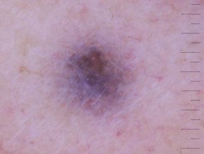
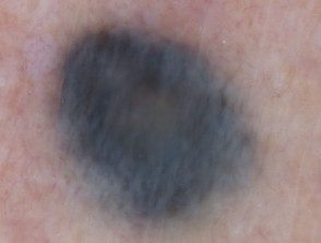
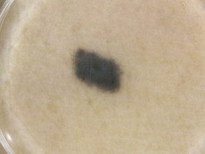
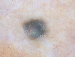
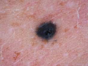
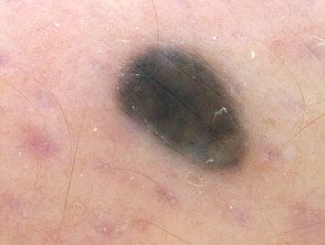
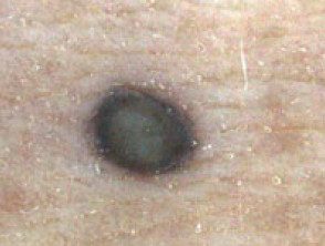
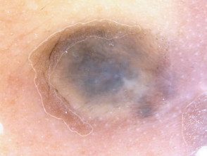
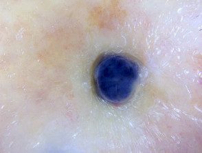
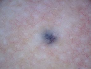
What are the dermoscopic features of a blue nevus?
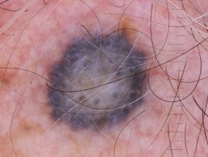
The dermoscopic mark of a blue nevus is homogeneous blue pigmentation. In some exceptional cases, they can present globules and blue points. [one].
The dermoscopic features of a blue nevus are:
- Seamless pattern
- Uniform blue color
- Lack of other structures.
- A well defined fade edge on outer skin.
- A diffuse Blue-white veil appearance over the entire surface.
The presence of any of the following structures should warrant further research for melanoma: net, dots, clods, stripes, glasses, additional colors.
Dermoscopic images of blue nevi
Blue nevus, cellular type
Blue nevus
Blue nevus
Blue nevus
Blue nevus
Blue nevus
Blue nevus
Nevus blue dermoscopy
Blue nevus
Blue nevus
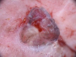
What is dermoscopy? differential diagnosis of a blue nevus?
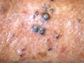
the primary The concern of an injury presenting as a blue nevus with an unknown history is that it could be one of the following:
- Nodular melanoma
- Melanoma metastasis
- Blue tattoo (eg, to indicate radiation site).
Differential diagnosis of blue nevus
Nodular melanoma
Nodular melanoma
Nodular melanoma
Melanoma metastasis
Melanoma metastasis
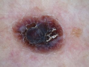
Dermoscopy of nodular melanoma.
Dermoscopy of nodular melanoma.
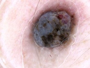
Dermoscopy of metastatic melanoma.
Dermoscopy of metastatic melanoma.
Radiation tattoo
Radiation Tattoo Dermoscopy
Which is the histological explanation of the characteristics of a blue nevus?
The histological explanation for a blue nevus is:
- Blue color: deep pigment in the lattice dermis
- Without structure: the heavy pigmented branched melanocytes are randomly arranged unrelated to the architecture of the overlying epidermis and thus appear unstructured in dermoscopy.
- Bluish white veil: orthokeratotic surface.
