
Lyme disease is a systemic infection caused by spirochete Borrelia burgdorferi. Chronic atrophic acrodermatitis is late cutaneous form of the disease
Histology Lyme disease
Biopsy of erythema Chronum migrans reveals a shallow and deep scarce infiltrate under an not involved epidermis (Figure 1). Sometimes there epidermal me dermal necrosis. High power examination reveals that the infiltrate is primarily lymphocytic (Figure 2). Often there is little eosinophils, mixed plasma cells and mast cells.
Chronic atrophic acrodermatitis lesions can show similar behavior. histopathology but the infiltrate is denser (figure 3). The infiltrator consists of lymphocytes and there are often numerous plasma cells (Figure 4) mixed with mast cells. There may be impressive superficial sclerosis and fibroplasia.
Lyme disease pathology
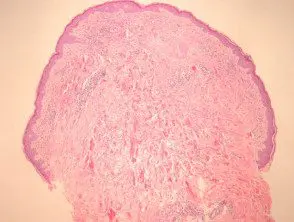
Figure 1
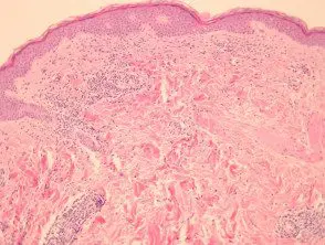
Figure 2
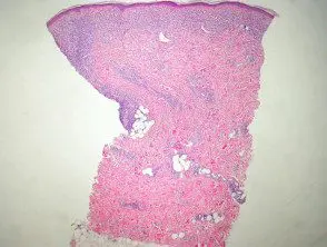
figure 3
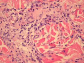
Figure 4
Special studies for Lyme disease.
Borrelia spirochetes can be identified with silver spots and immunohistochemical methods in some cases. Many cases will be negative with these methods. Polymerase chain reaction (PCR) can also be used.
Differential diagnosis Lyme disease pathology
Insect sting reaction: Chronic migratory erythema lesions can be nonspecific and accurately mimic an insect sting reaction. This is particularly problematic when silver spots and immunohistochemical studies are negative. Clinical correlation, PCR studies, and serological correlation may be required.
Sclerosing Disorders: Chronic atrophic acrodermatitis can cause a sclerosing reaction to closely simulate lichen sclerosus or eosinophilic fasciitis
