

Ad
Skin cancer
Application to facilitate skin self-examination and early detection. read more.
What is a seborrheic keratosis?
Seborrheic keratosis is harmless. warty spot that appears during adult life as a common sign skin aging. Some people have hundreds of them.
Seborrheic keratosis (or seborrheic keratosis, using American spelling) is also called SK, basal cell papilloma, senile wart, brown wart, wisdom wart or barnacle. The descriptive term, benign Keratosis is a broader term used to include the following related scaly skin lesions:
- Seborrheic keratosis
-
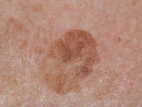
Solar lentigo (which can be difficult to distinguish from flat seborrheic keratosis)
-
Lichen planus type keratosis (arising from a seborrheic keratosis or a solar lentigo).
Seborrheic keratosis
Seborrheic keratosis
Seborrheic keratosis
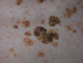
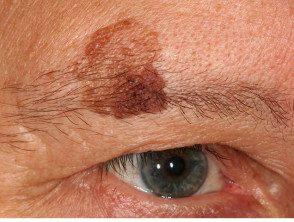
Pigmented seborrheic keratosis
Who gets seborrheic? keratosis?
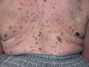
Seborrheic keratoses are extremely common. It has been estimated that more than 90% of adults over 60 have one or more of them. They occur in men and women of all races, usually beginning to erupt in the 1930s or 1940s. They are rare under the age of 20.
What causes seborrheic keratoses?
The exact cause of seborrheic keratoses is not known.
The name is misleading, because they are not limited to a seborrheic distribution (scalp, mid-face, chest, upper back) as in seborrheic dermatitis, nor are they formed from sebaceous glands, such as sebaceous hyperplasianor are they associated with tallow - Which is greasy.
Seborrheic keratoses are considered degenerative in nature. As time passes, seborrheic keratoses become more numerous. Some people inherit a tendency to develop a large number of them. The researchers have noted:
- Eruptive seborrheic keratoses can follow sunburn or dermatitis.
- Friction of the skin may be the reason why they appear in the folds of the body.
-
The viral cause (eg, Human Papillomavirus) seems unlikely.
- Stable and clone mutations o activation of FRFR3, PIK3CA, RAS, AKT1 and EGFR genes They are found in seborrheic keratoses.
- Seborrheic keratosis can arise from solar lentigo.
- FRFR3 mutations also arise in solar lentigines. These mutations are associated with increased age and location in the head and neck, suggesting a role for Ultraviolet radiation in these injuries
- Seborrheic keratoses do not harbor tumor suppressor gene mutations
- Epidermal growth factor receiver inhibitors (used to treat Cancer) often result in an increase in keratosis warts (warts).
What are the clinical characteristics of seborrheic keratoses?
Seborrheic keratoses can arise on any area of the skin, covered or uncovered, with the exception of the palms and soles of the feet. They do not arise from mucous membranes
Seborrheic keratoses have a highly variable appearance.
- Flat or elevated papule or license plate
- 1 mm to several cm in diameter
- Skin color, yellow, gray, light brown, dark brown, black or mixed colors.
- Smooth, waxy or warty surface
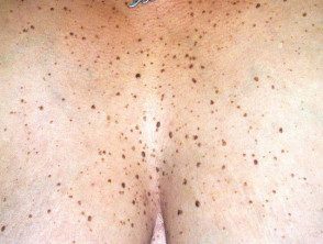
- Solitaire or grouped in certain areas, such as inside the scalp, under the breasts, over the spine, or in the groin
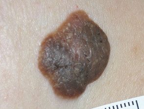
They seem to cling to the skin's surface like barnacles.
Seborrheic keratosis
Seborrheic keratosis
Seborrheic keratosis
Seborrheic keratosis
- See more images of seborrheic keratosis
- See dermoscopic images of seborrheic keratoses
Variants of seborrheic keratoses
Variants of seborrheic keratoses include:
-
Lentigo solar: flat circumscribed pigmented patches in places exposed to the sun
- Skin disease papulosa nigra: small, pedunculated and highly pigmented seborrheic keratoses on the head and neck of individuals with dark skin
- Keratosis stucco: gray, white or yellow papules in the lower extremities
- Invested follicular keratosis
- Large cell acanthoma
- Lichenoid keratosis: a inflammatory phase prior to the involution of some seborrheic keratoses and solar lentigines.
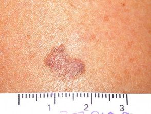
Benign keratosis
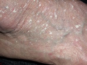
Stucco keratosis
Papulous nigra dermatosis
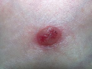
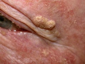
Irritated seborrheic keratosis
Lichenoid keratosis
Complications of seborrheic keratoses
Seborrheic keratoses are not premalignant Tumors However:
-
Skin cancers are sometimes difficult to distinguish from seborrheic keratoses.
- Skin cancer can arise by chance or collide with seborrheic keratosis.
Very rarely, eruptive seborrheic keratoses may denote an underlying internal malignancy, most often gastric adenocarcinoma. the paraneoplastic syndrome It is known as the sign of Leser-Trélat. Eruptive seborrheic keratoses that are not associated with cancer are sometimes described with a Leser-Trélat pseudo-sign.
Eruptive and irritated seborrheic keratoses can also arise as a adverse reaction to a medicine, such as adalimumab, vemurafenib, dabrafenib, 5-fluorouracil, and many chemotherapy drugs
An irritated seborrheic keratosis is inflammation, redness, and scabs. injury. It can lead to eczematous dermatitis around seborrheic keratosis. Dermatitis can also trigger the appearance of new seborrheic keratoses.
How is seborrheic keratosis diagnosed?
The diagnosis of seborrheic keratosis is often easy.
- A pasted, well-demarcated warty plaque
- Other similar injuries
Sometimes seborrheic keratosis can resemble skin cancer, like basal cells. carcinoma, scaly cell carcinoma or melanoma.
Dermoscopy often shows a disordered structure in seborrheic keratosis, as is also true for skin cancer. Dermoscopic diagnostic clues exist for seborrheic keratosis, such as multiple orange or brown lumps (due to curb in cracks in the skin surface), white milia-like clods, and thick curved ridges and grooves that form a cerebral or cerebriform pattern.
If doubt persists, a seborrheic keratosis may suffer a partial shave or a blow biopsy or diagnosis excision.
The dominant histopathological The characteristics of seborrheic keratosis can be described as:
- Melanoacanthoma (deeply pigmented)
- Acanthotic
- Hyperkeratotic or papillomatous
- Adenoids or lattice
- Clonal or nested
- Adamantinoid or mucinous
- Desmoplastic
- Irritated.
What is the treatment for seborrheic keratoses?
An individual seborrheic keratosis can be easily removed if desired. The reasons for removal may be that it is unsightly, itches or catches clothing.
The methods used to remove seborrheic keratoses include:
- Cryotherapy (liquid nitrogen) for thinner injuries (repeated if necessary)
-
Curettage and / or electrocautery
- Ablative To be surgery
- Shaving biopsy (shaving with a scalpel)
- Focal chemical peel with trichloroacetic acid
All methods have disadvantages. Treatment-induced loss of pigmentation It is a particular problem for dark skinned patients. There is no easy way to remove multiple injuries in one go.
How can seborrheic keratoses be prevented?
It is unknown how to prevent seborrheic keratoses.
What is the prognosis for seborrheic keratoses?
Seborrheic keratoses tend to persist. Occasionally, single or multiple lesions may remit spontaneously or through the mechanism of lichenoid keratosis.
Those associated with dermatitis may regress after it has been controlled.
