
What is a dental? breast?
A dental sinus is an abnormal canal that drains from a long-standing dental tooth. abscess associated with a necrotic or dead tooth. A dental sinus can drain to:
- the inside of the mouth (an intraoral sinus) or
- The surface of the skin of the face or neck (an extraoral or orofacial sinus).
Intraoral sinuses are the most common form and most necrotic teeth have been reported to drain this way.
Who has a dental sinus?
A dental sinus usually results from a chronic infection in long-standing necrotic dental pulp (a dead tooth). Tooth decay is usually due to tooth decay or trauma. Cavities occur due to poor dental hygiene and regular consumption of refined sugars. Other predisposing factors for tooth decay include:
- removable dentures (due to food stagnation)
- xerostomia (dry mouth: saliva protects against infection).
Infection is more likely after endodontic work and in patients who are immunosuppressed, having chemotherapy or suffering from blood dyscrasias.
The direction a sinus takes within the mouth or towards the skin determines which tooth it is involved in and follows the path of least resistance: the thickness of the bone, as well as the muscle attachments and fascial planes direct the route of drainage.
Intraoral sinuses generally occur in the groove on the side of the cheek near the tip of the involved tooth.
Most extraoral sinuses start from a tooth in the lower jaw and drain towards the chin or below the chin or jaw line (submental or submandibular area). Those that originate from a tooth in the upper jaw can drain down to the cheek or near the nose. The site of an extraoral sinus opening is often located a great distance from the infected tooth.
What are the clinical characteristics of a dental sinus?
Infected necrotic pulp can cause severe toothache before sinus or fistula The disappearance of pain without dental treatment develops, it can be an important clue that the abscess has drained and formed a sinus. However, the process can also occur painlessly.
The intraoral dental sinus may appear as a persistent mouth ulcer It drains pus, causing a bad taste in the mouth. The extraoral dental sinus may present as a persistent, draining sore or as a lump on the face. It is usually painless. the download it can be pus or blood stained. The sinus opening can be seen with careful examination.
Because toothache is generally absent, the patient frequently presents to a physician rather than a dentist. As the extraoral dental sinus is a rare condition, it is often initially misdiagnosed as a more common skin condition, such as a skin Cancer, boil or other skin infection, pyogenic granuloma, trauma, foreign body or other granuloma, cyst or one of the other breast forms for the face and neck and fistulas.
Reappearance despite antibiotics or surgery is a clue to the correct diagnosis.
An obviously decayed tooth in the mouth or a history of deep filling usually suggests which tooth is causing the injury. The relevant tooth may be discolored or tender when touched. There may be evidence of previous dental or endodontic work or poor oral hygiene in general.
However, a tooth can fill for many years before dying without pain, and thus examining the teeth clinically may not show any obvious abnormalities. The tooth may not respond to the cold or electric pulp test (pulp vitality test / pulp sensitivity test).
Dental abscesses can also be complicated by osteomyelitis (bone infection), cellulitis (redness, swelling), or a facial abscess. Head or neck lymph nodes it can become larger It is very important to quickly treat facial swelling as the infection can spread to other parts of the body or endanger the airways.
Extraoral dental sinus
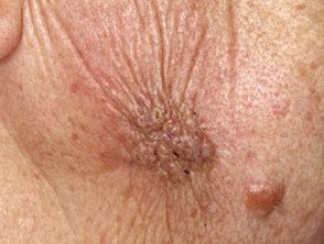
Dental sinus
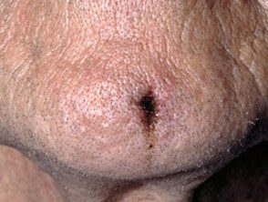
Dental sinus
How is a dental sinus diagnosed?
The clinical clues should be:
- Previous history of toothache, trauma to the face, or deep filling
- A persistent drain injury in the mouth or on the face or neck often despite repeated courses of antibiotics and / or surgery
- Signs in dental examination.
Radiology (X-rays) is the most important investigation, as it usually shows an area of bone loss around the root tip of the chronically infected tooth. When the involved tooth is not obvious, a gutta-percha (gum) point can be inserted into the sinus to follow its course to the relevant tooth. Rarely Connecticut scan or Magnetic resonance it is required.
If possible, surgery should be avoided as it will not solve the problem and can lead to unnecessary scarring. Biopsies (if taken) may be reported to show abscess, granuloma or a epitheliumscratched tract.
A variety of bacteria can be isolated from a swab strictly including anaerobic gram negative rods and aerobic gram-positive cocci.
What is the treatment for a dental sinus?
Removal of the entire tooth (extraction) or necrotic dental pulp (root canal / root canal treatment) is the only successful treatment for a dental sinus.
Antibiotics such as penicillin or metronidazole may also be required.
The sinus will usually heal 1–2 weeks after successful root canal extraction or treatment. There may be residual scars if biopsies or surgery were done. Otherwise, there could be a slight dimple or a change in the color of the skin surface that generally improves with time.
