
What is a blistering disease?
A blistering disease is a condition in which there are fluid-filled skin lesions.
- Vesicles They are small blisters less than 5 mm in diameter.
- A bulla It is a larger blister. Note that the plural of bulla is bullas.
- The blisters may break or the top of the blister may come off forming a erosion. Oozing serous fluid shapes Cortex.
Blistering diseases
Vesicles
A noise
Cortex
Acute blistering diseases
Acute blistering diseases can be generalized or located to a body site and are due to infection or inflammatory disorders although more commonly eczematous, widespread acute blisters can be life threatening and often require hospitalization.
Acute blistering conditions should be investigated by taking swabs to bacterial and viral culture. A skin biopsy It can be useful to make a diagnosis.
Acute generalized blistering diseases
Acute febrile neutrophilic skin disease
- Neck, limbs, upper trunk
- Pseudovesicular platesblisters pustules, purpleor ulceration
- Disease associations: rheumatoid arthritisInflammatory bowel disease autoimmune arthritis, myeloid dysplasia
- Suggestive biopsy
Atypical enterovirus infection
- Extended vesicular eruption
- It clears in a few days.
- When it affects areas of atopic eczemaThe enteroviral infection is called eczema coxsackium.
Chicken pox / chicken pox
- Childhood illness; more severe in adults
- Scalp, face, oral mucous membrane, trunk
- Culture/PCR Herpes chickenpox zoster
Acute febrile neutrophilic dermatosis.

Atypical enterovirus infection
Chicken pox / chicken pox
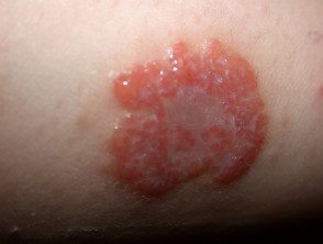
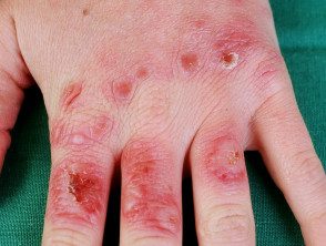
Herpetic eczema
- Atopic history dermatitis
- Monomorphic group of umbilical vesicles
- Herpes simplex culture / PCR
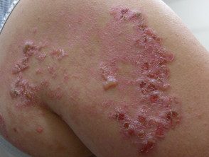
Dermatitis
- Atopic dermatitis
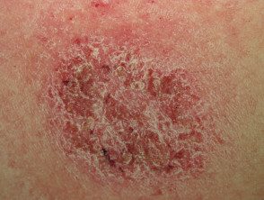
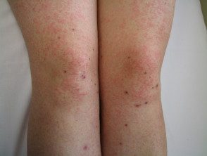
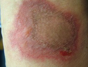
- Discoid eczema
Discoid eczema
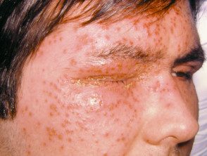
Herpetic eczema
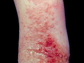
Atopic dermatitis
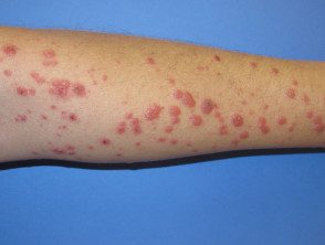
Polymorphic light eruption
- It affects the sites of the body exposed to the sun (hands, upper chest, feet
- Papulesplates sometimes targettoid
- May have spare face
- Arises within hours of exposure to bright sunlight
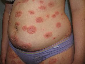
Erythema multiform
- A reaction, for example, to an infection.
- An acute rash of papules, plaques, target lesions
- Acral distribution: cheeks, elbows, knees, hands, feet
- Can have mucositis (lips, conjunctivagenitals)
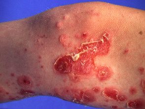
Stevens-Johnson syndrome / / toxic epidermal necrolysis
- Very bad patient
- Mucous membrane intervention
- Almost always drug induced
- Rarely due to mycoplasma infection
- Painful red skin may peel off sheets or have multiple uniting blisters

Polymorphic light eruption
Erythema multiforme
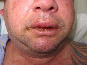
Stevens Johnson Syndrome / Toxic Epidermal Necrolysis
Drug hypersensitivity syndrome
- Medication started up to 8 weeks before start
- Morbid blistering rash (without necrolysis)
- Often mucosal involvement
- Multi-organ damage (renal, hepaticrespiratory hematological)
- Often marked eosinophilia
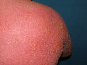
Staphylococcal scalded skin syndrome
- Little boy
- Miserable
- Red skin comes out on sheets
- Evidence of staphylococcal infection
Drug hypersensitivity syndrome
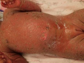
Staphylococcal scalded skin syndrome
Acute localized blistering diseases
Acute dermatitis
- Contact dermatitis
- Plant dermatitis
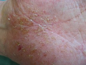
- Pompholyx
Bullous impetigo
- Quickly expanding license plate
- Staphylococcus aureus swab
-
Wound infections, scabies, etc.
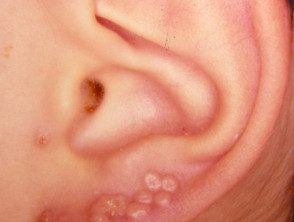
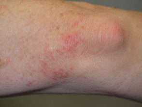
Chilblains
- Toes
- Exposed to cold
- Purple tender plates
Plant dermatitis
Blistering impetigo
Chilblains
Enteroviral vesicular stomatitis
- Hand, foot and mouth disease
It clears in a few days.
Erysipelas
- Acute febrile illness
- Streptococcus pyogenes swabs
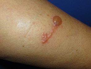
Fixed drug eruption
- Newspaper eruption, often in the same place
- Due to an intermittent medication that has been taken within 24 hours of the rash onset
- Single or few injuries
- Central blister
Enteroviral vesicular stomatitis
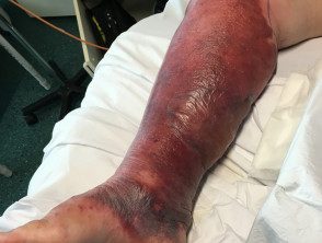
Erysipelas
Fixed drug eruption
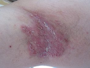
Herpes Simplex
- Monomorphic, umbilicated
- Herpes simplex culture / PCR
Herpes zoster (shingles)
- Dermatomic
- Culture / PCR Herpes zoster
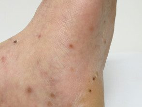
Insect bites and stings
- Crops of Urticated papules
- Central vesicle or punctum
- Favor exposed sites
- Blisters due to contact with various beetles (eg, dermatitis padarus, lax beetle dermatosis) and caterpillars
Herpes Simplex
Herpes infection
Insect bites and stings
Miliaria
- Central trunk
- Sweat rash
- The vesicles are very shallow
Necrotizing fasciitis
- Very ill; septic shock
- Rapid spread of cellulite with purpura / blisters.
- Anesthetic areas in early lesions
-
Essential bacterial culture
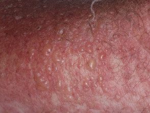
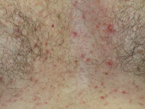
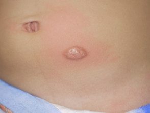
Transient acantholytic skin disease
- Sharp or chronic
- Old man
- Itching or asymptomatic
- Crusted papules
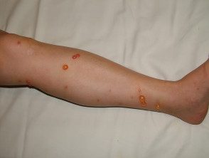
Trauma
- Injury history or neuropathy
- Friction, thermal burns, Ultraviolet radiation sunburn, chemical burns, fractures
Miliaria
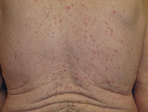
Transient Acantolytic Dermatosis
Trauma-ultraviolet radiation
Chronic blisters
Diagnosis of chronic blisters often requires a skin biopsy to histopathology and direct immunofluorescence. A blood test to antibodies (indirect immunofluorescence) can also be helpful in making the diagnosis of an immunobullous disease.
Burning genodermatosis
Epidermolysis bullosa
- Various types
- Start at birth or early childhood
Fogo selvagem
- Also called endemic foliaceous pemphigus
- Onset in childhood or adolescence
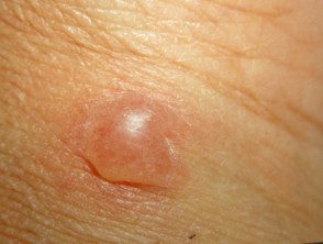
Mastocytosis
- Various types
- Often start in childhood
Benign family pemphigus
- Also called Hailey-Hailey disease
- Confined to push-ups
Genodermatosis blisters
Epidermolysis bullosa
Mastocytoma
Benign family pemphigus
Chronic blisters acquired
Bullous systemic lupus erythematosus
- The patient has systemic lupus erythematosus.
- Subepidermal bullae
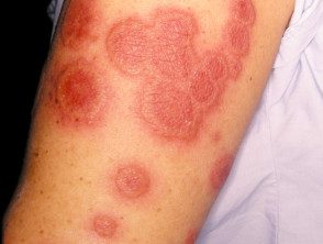
Bullous pemphigoid
- Mainly cutaneous (rarely mucous)
- Mainly affects the elderly (rarely infants, children)
- Often associated stroke or dementia
- Subepidermal bullae
- Eczematous or urticarial pioneers
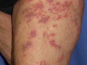
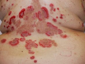
Herpetiform dermatitis
- Associated gluten associated enteropathy
- Intense itching the vesicles are often removed by scratching leaving erosions
- Symmetrical on scalp, shoulders, elbows, knees, buttocks
Chronic blisters acquired
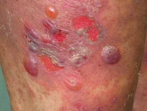
Bullous pemphigoid
Herpetiform dermatitis
Other immunobullous diseases
- Cicatricial pemphigoid
-
Gestational pemphigoid
- Linear IgA dermatosis
- Acquired epidermolysis bullosa
-
Pemphigus vulgaris
- Leaf Pemphigus
- Paraneoplastic pemphigus
-
Drug-induced pemphigus
- Pemphigus IgA
- Ocular involvement in autoimmune blisters
Cutaneous porphyria delays
- Metabolic photosensitivity
- Skin fragilitybullas milia
- Back of hands, face
- Onset in middle age
Other immunobullous diseases
Gestational pemphigoid
Pemphigus vulgaris
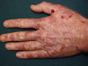
Cutaneous porphyria delays
