
What is the white sponge? nevus?
White sponge nevus (Mendelian inheritance in man [MIM] number 193900) is a rare genetic condition causing fluffy white lesions of the mucous membranes, most commonly the mouth. It has also been called congenital leukokeratosis mucous membrane oris hereditary leukokeratosis, white folded gingivostomatitis, oral epithelial nevus and oral white sponge nevus.
White sponge nevus
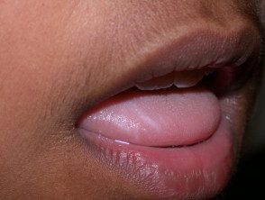
White sponge nevus
Who receives the white sponge nevus?
White sponge naevus is a autosomal Dominant condition, which means that the children of an affected parent have a 50:50 chance of inheriting the condition. However, it can occasionally be sporadic with no history of affected family members.
It is believed to be rare, but it may be underdiagnosed, especially in the absence of similar injuries affecting family members.
The degree of expression varies from case to case, from absence to slight to extensive changes. Some authors claim that women are more commonly affected than men.
White sponge nevus may be present at birth or appear later in childhood or adolescence.
Clinical characteristics of the white sponge nevus
White sponge naevus presents as bilateral, sometimes symmetricalSoft, white, raised lesions of mucous membranes. The surface may appear bent and feel spongy, not hard. It cannot be separated. The change can be quite subtle and located or it may involve the entire inside of the mouth.
It usually does not cause any symptoms, but patients may complain of roughness and appearance.
The mouth is the most common affected site and the inside of the cheeks is the most common site within the mouth. However, the mucous membranes inside the nose, the esophagus, the genitals (vulva and vagina) and anorectal sites may also be involved. Usually the mouth is the first place noticed.
No changes are noted in other parts of the skin, nail, hair o Teeth This is important when considering other possible causes of injury.
There have been no oral reports Cancer developing on a white sponge nevus.
How is white sponge nevus diagnosed?
The combination of the clinical appearance, the absence of other skin problems and a positive family history should increase the possibility of this diagnosis, which is later confirmed in biopsy of the injury and pathology exam. the histopathology of white spongy nevus is very characteristic and in particular shows large areas of large cells with light skin in the epidermis.
Diagnosis can now be made definitively in genetic analysis and identification of a mutation in a highly preserved sequence of a copy of the gene coding for the curb 4 or 13 proteins.
Many other genetic conditions can show white spots in the mouth, but they can usually be excluded on clinical examination, as they all have visible changes in other parts of the skin, nails, etc. These include pachyonychia congenital, Darier's disease, dyskeratosis congenital and hereditary benign intraepithelial dyskeratosis.
Most often, white sponge nevus is misdiagnosed as oral yeast infection (thrush), but this may be excluded in microbiological swabs, lack of response to antifungal therapy, and biopsy. One clue in the clinical examination is that the white patches of yeast infection may peel off.
The second most common misdiagnosis is oral lichen planus and this is readily distinguishable on biopsy.
Other causes of localized white spots in the mouth that can cause confusion include leukoplakia, chewing on the cheeks, chewing tobacco or betel nut (oral submucosa fibrosis), syphilis and lupus erythematosus. Again, the pathology of a biopsy will clarify this.
Naeuvs white sponge treatment
One of the important reasons for making this diagnosis is to avoid unnecessary treatment. In general, explanation and reassurance are all that is required.
When requesting treatment, there have been some reports of success control using current or systemic antibiotics
-
Tetracycline mouthwash - 0.25% aqueous tetracycline solution, 5 ml twice a day.
-
oral tetracycline: initially 250 mg, four times a day for 4 weeks, followed by 250 mg once a week to maintain response
-
oral amoxicillin: initially 250 mg three times a day for 4 weeks, followed by 250 mg once a week to maintain improvement.
-
erythromycin may help if tetracycline and amoxicillin are inappropriate due to allergy.
These are not curative, but can sometimes induce a complete or partial response. remission which can be maintained by continuing with a low dose. When the antibiotic is stopped, the condition recurs, usually after weeks or months. It is not clear why these antibiotics should help some patients.
Many other treatments have been tried without success, including topical antifungal agents, oral antifungal therapies, retinoids like acitretin and isotretinoin, vitamins and other antibiotics like metronidazole and trimethoprim + sulfamethoxazole.
Proposed mechanism for the development of white sponge nevi
It is now known that the white sponge nevus is due to a mutation in the highly conserved regions of domain 1a or 2b of the genes keratin 4 or 13 encoding. Most commonly a nonsense mutation is found that results in a change in a amino acids that interferes with the assembly of the intermediate filaments. Keratin proteins 4 and 13 are only produced orally, nasal, esophageal and anogenital mucous membranes, hence the reason for the change that occurs in these areas. Electron microscopy white sponge nevus shows the abnormal aggregation of the intermediate filaments in the large clear cells.
