
Introduction
Cutaneous Plasmacytoma is a rare manifestation (<1%) of multiple myeloma or plasma cell leukemia. Disproportionately affects IgA and IgD proliferations and is associated with worsening forecast.
Histology plasmacytoma
There is a dense cellular infiltrate (Figure 1) filling the biopsy sample. This is composed of a monotonic population of plasma cells (Figure 2).
There are histological classification criteria described by Bartl et al in 1987. Cases can also be classified differentiated (grade I), moderately differentiated (grade II) or poorly differentiated (grade III) based on plasma cells morphology.
Pathology of cutaneous plasmacytoma.
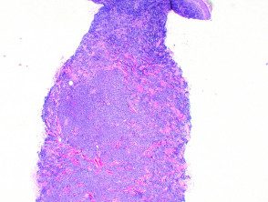
Figure 1
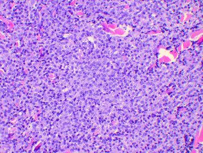
Figure 2
Special spots in plasmacytoma
Plasma cells show a monoclonal profile confirmed by increased kappa or lambda staining in immunohistochemistry (figures 3, 4) or FISH. Plasma cells are also highlighted by CD138 immunostaining (Figure 5).
Special plasmacytoma stains
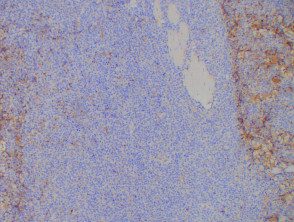
figure 3
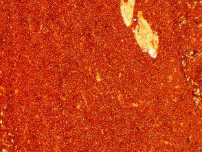
Figure 4
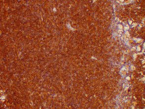
Figure 5
