
What is impetigo?
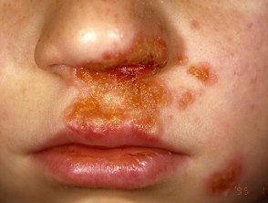
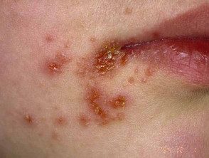
Impetigo is common acute superficial bacterial skin infection (pyoderma) It is characterized by pustules and honey-colored scab erosions ('school sores').
The term impetiginization It is used for secondary superficial infection of a wound or other skin condition. Ulcerated impetigo is called an ecthyma.
What causes impetigo?
Impetigo is usually caused by Staphylococcus aureus. Not-bullous Impetigo can also be caused by group A beta-hemolytic streptococcusStreptococcus pyogenes)
Non-blistering impetigo
In non-bullous impetigo, staphylococci and streptococci invade a minor site. trauma where exposed proteins allow bacteria to adhere.
Ecthyma
The ecthyma is usually due to Streptococcus pyogenes, but coinfection with Staphylococcus aureus it can happen.
Blistering impetigo
Blistering impetigo is due to a staphylococcal peel toxins (exfoliatina A - D), what objective desmoglein 1 (a desmosomal accession glycoprotein) and divide the superficial epidermis through granular layer. No trauma is required, as the bacteria can infect intact skin.
Who has impetigo?
Impetigo is more common in children (especially children), but it can also affect adults if they have low immunity to the bacteria. This predominant Worldwide. The maximum onset is during the summer, and is more frequent in developing countries.
The following factors predispose to impetigo
- Atopic eczema
- Scabies
- Skin trauma: chickenpox, insect bite, abrasion, laceration, thermal burn, dermatitissurgical wound
What are the clinical characteristics of impetigo?
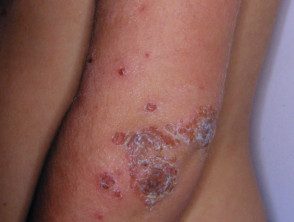
Primary impetigo mainly affects exposed areas such as the face and hands, but can also affect the trunk, perineum and other places on the body. Presents with single or multiple irregular crops with irritable surface plates. These spread as they heal, forming cancel or arcuate Injuries
Although many children are fine otherwise, lymphadenopathymild fever and discomfort it can happen.
Non-blistering impetigo
Non-blistering impetigo starts like a rose taint which evolves to a vesicle or pustule and then in crusted erosions. Untreated impetigo usually resolves in 2 to 4 weeks without scarring.
Ecthyma
The ecthyma begins as nonblistering impetigo, but develops into a perforation. necrotic ulcer which heals slowly, leaving a scar.
Blistering impetigo
Blistering impetigo presents with small vesicles that evolve in flaccid transparent bullas. Heals without scarring.
Impetigo
Impetigo
Impetigo
Impetigo
See more images of impetigo.
Impetigo complications
Soft fabric infection
The bacteria that cause impetigo can become invader, which leads to cellulite and lymphangitis; subsequent bacteremia could result in osteomyelitis, septic arthritis or pneumonia
Staphylococcal scalded skin syndrome
In infants under six years of age or adults with renal insufficiency, located Blistering impetigo due to specific staphylococcal serotypes can lead to a sick child with generalized Staphylococcal scalded skin syndrome (SSSS). Superficial crust then tender cutaneous denudation on the face, in push-ups, and elsewhere it is due to circulating exfoliatin / epidermolysin, rather than a direct skin infection. Leaves no scar.
Toxic shock syndrome
Toxic shock syndrome is rare and rarely preceded by impetigo. It causes fever diffuse erythematous then flaking eruption, hypotension and involvement of other organs.
Post-streptococcal glomerulonephritis
Group A streptococcal infection can rarely lead to acute post-streptococcal glomerulonephritis 3 to 6 weeks after skin infection. Associates with anti-DNase B and antistreptolysin O (ASO) antibodies.
Rheumatic fever
Group A streptococcal skin infections have rarely been associated with cases of rheumatic fever and rheumatic heart disease. This is believed to occur because the strains of group A streptococci generally found on the skin have moved to the throat (the most common site for infection associated with rheumatic fever).
How is impetigo diagnosed?
Impetigo is usually diagnosed clinically, but can be confirmed with bacterial swabs sent for microscopy (Gram-positive cocci are observed), culture and sensitivity.
A blood count can reveal neutrophils leukocytosis when impetigo is extended.
Skin biopsy It is rarely necessary. the histological The characteristics of impetigo are characteristic.
Non-blistering impetigo
- Gram-positive cocci
- Intraepidermal neutrophilic pustules
- Dense inflammatory infiltrate on top dermis
Blistering impetigo
- Separate through the granular layer of the epidermis without inflammation or bacteria
- Acantolytic cells
- Minimal inflammatory infiltrate in the upper dermis
- It resembles pemphigus foliaceus
Ecthyma
- Full thickness skin ulceration
- Gram stain shows cocci within the dermis
What is the treatment for impetigo?
The following steps are used to treat impetigo. [1,2].
- Clean the wound; use wet soaks to gently remove scabs.
- Apply antiseptic 2–3 times a day for five days (povidone iodine, hydrogen peroxide to 1% cream, chlorhexidine, superoxidized solution and others).
- Cover the affected areas.
Oral antibiotics are recommended if:
- Symptoms are significant or severe (fever, malaise)
- There are more than three injuries.
- There is a high risk of complications.
- The infection is not resolving or is unlikely to resolve.
Suitable oral antibiotics in New Zealand are flucloxacillin 500 mg four times daily for 5 days (adult dose), and in case of allergy or bacterial resistance, trimethoprim + sulfamethoxazole 960 mg, twice daily, for five days (adult dose), erythromycin 800 mg twice daily for 5 days, or cephalexin 1 g twice daily for 5 days [2].
In New Zealand, antibiotic. ointment (fusidic acid, mupirocin or retapamulin) is not recommended, due to the potential to develop bacterial resistance and allergic contact dermatitis.
To prevent reappearance:
- Try carrier sites: apply antiseptic ointment to the nostrils
- Wash daily with antibacterial soap or soak in a bleach bath
- Cut nail and keep your hands clean
- Identify and treat the source of the reinfection, usually another infected person or carrier in the home.
To reduce the chance of passing the infection to another person:
- Avoid close contact with others.
- Children should be kept away from school until the scabs have dried or for 24 hours after starting oral antibiotics.
- Use separate towels and flannels
- Change and wash clothes and clothing every day.
