
Leiomyosarcoma is a evil one tumor smooth muscle Rarely affects the skin.
Histology leiomyosarcoma
The scanning power of leiomyosarcoma histology shows poor circumscribed tumor nodule What can be dermal based on less common dermal leiomyosarcoma (Figure 1) or deeply infiltrating the subcutaneous to form. The tumor is made up of a spindle cell proliferation forming rough packets and fascicles (Figures 2 and 3). High power shows spindle cells shaped like a cigar nuclei with prominent cytology atypia and mitotic figures (Figures 4 and 5).
Leiomyosarcoma pathology
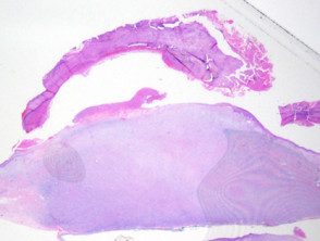
Figure 1
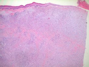
Figure 2
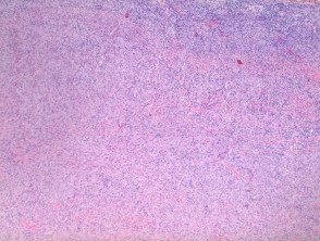
figure 3
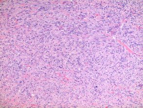
Figure 4
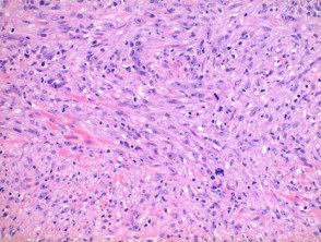
Figure 5
Special spots in leiomyosarcoma
Immunoperoxidase staining is positive for muscle markers SMA (smooth muscle actin), HHF35 (pan muscle actin), h-caldesmon, and Desmin. Desmin negative staining is seen infrequently. A rare number of cases have shown cytokeratin or EMA (epithelial membrane antigen) staining.
Histological leiomyosarcoma variants
Atypical leiomyoma: this injury it probably falls on a spectrum between leiomyoma and leiomyosarcoma. Reliable criteria to exclude malignancy in cutaneous Leiomyosarcomas are missing, but reported uterine discrimination characteristics include: size less than 5.5 cm, mitotic count less than 7 per 10 high-power fields in the presence of nuclear atypia and lack of tumor cells necrosis.
Se usa un leiomyoma mitótico activo para describir una lesión que carece de atipia nuclear significativa o necrosis tumoral, pero con entre 5 y 15 mitosis per high power field.
Differential diagnosis leiomyosarcoma
Other malignant spindle cell tumors to consider include the spindle cell. scaly cell carcinomaevil one peripheral nerve sheath tumor, spindle cell melanoma, angiosarcoma or atypical fibroxanthoma. Immunoperoxidase staining is generally required to differentiate between these lesions.
