

Ad
Skin cancer
Application to facilitate skin self-examination and early detection. read more.
What is it acral lentiginous melanoma?
Acral lentiginous melanoma is a type of melanoma that arises on the palms or soles. It is a form of melanoma that is characterized by its site of origin: palm, sole of the foot or under the nail (subungual melanoma). It is more common in the feet than the hands. may arise de novo in normal-appearing skin, or may develop within existing skin melanocytic nevus (Mole).
Acral lentiginous melanoma begins as a slow growing plane patch of discolored skin. At first, the evil one The cells remain within the tissue of origin, the epidermis - the in the place phase of melanoma, which can persist for months or years.
Acral lentiginous melanoma develops invader when melanoma cells cross the basement membrane of the epidermis, and malignant cells enter the dermis. Rapid growth nodular Melanoma can also arise within acral lentiginous melanoma and proliferate deeper within the skin.
Who gets acral lentiginous melanoma?
About 1 to 3% melanomas in Australia and New Zealand they are acral lentiginous melanoma. Melanoma is a potentially serious skin condition. Cancer that arises from pigment cells (melanocytes) Although acral lentiginous melanoma is rare in Caucasians and people with darker skin types, it is the most common subtype in people with darker skin types.
Acral lentiginous melanoma is relatively rare compared to other types of melanoma. There is no connection to skin color (skin phototype), and it occurs at the same rate in white, brown or black skin. Acral lentiginous melanoma accounts for the 29-72% of melanoma in dark-skinned individuals, but less than the 1% of melanoma in light-skinned individuals, as they are prone to the more common types of sun-induced melanoma, such as spreading melanoma. superficial and lentigo malignant melanoma
Acral lentiginous melanoma is equally common in men and women. The majority arises in people over 40 years of age.
The cause or causes of acral lentiginous melanoma are unknown. It is not related to sun exposure.
What is acral lentiginous melanoma like?
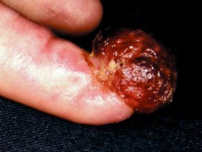
Acral lentiginous melanoma on the palm of the hand, soles of the feet, or toes begins as an enlarged patch of discolored skin. It is often thought at first to be a stain. It can also be amelanotic (no-pigmented, usually red). Like other flat forms of melanoma, it can be recognized by the ABCDE rule: asymmetry, irregularity of the border, color variation, large diameter and evolution.
Features of acral lentiginous melanoma include:
- Large size: >6 mm and often several centimeters or more in diameter at diagnosis
- Variable pigmentation: most often a mixture of brown, blue-gray, black and red colors
- A smooth surface at first, then becomes thicker with an uneven surface that may be dry or warty
- Ulceration or bleeding
Acral lentiginous melanoma
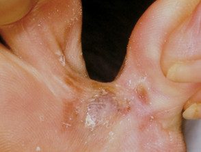
Acral lentiginous melanoma
Acral lentiginous melanoma
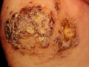
Acral lentiginous melanoma
See more images of acral lentiginous melanoma.
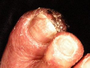
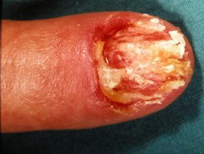
Subungual melanoma
When acral lentiginous melanoma arises in the nail region, it is known as nail unit melanoma. If it starts in the womb (nail growth area), it is called subungual melanoma. It can be presented as diffuse discoloration or irregular pigmented longitudinal bands on the nail plate. Advanced melanoma destroys the nail plate completely. It may be amelanotic.
Melanoma of the nail unit
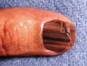
Subungual melanoma
Subungual melanoma
Subungual melanoma
See more images of melanoma of the nail unit.
What is the cause of acral lentiginous melanoma?
Acral lentiginous melanoma is due to the development of malignant pigment cells (melanocytes) along the basal cap of the epidermis. These cells may arise from an existing melanocytic nevus or, more often, from normal-appearing skin. It is unknown what causes melanocytes to become malignant, but it involves genetic mutations.
What tests should be done?
Dermoscopy
Dermatoscopy, or the use of a dermatoscope, by a dermatologist or another doctor trained in its use, can be very useful in distinguishing acral lentiginous melanoma from other skin lesions, in particular:
- Melanocytic naevi (moles)
- Viral warts
- Bleeding – subcorneal hemorrhage or subungual hematoma
The most frequently observed dermoscopic features of acral lentiginous melanoma are:
- Asymmetric structure and colors
- Pigment Parallel Ridge Pattern distribution
- Blue-gray structures.
See dermoscopy of acral lentiginous melanoma.
Biopsy
If the skin injury suspected acral lentiginous melanoma, it is better to cut it (excision biopsy). It is best to avoid a partial biopsy, except in unusually large lesions. An incisional or puncture biopsy could miss a focus of melanoma arising in a preexisting nevus. However, sometimes the lesion is very large and before significant surgery is performed, a biopsy is arranged to confirm the diagnosis. Biopsy of a lesion suspicious for acral lentiginous melanoma should remove a long ellipse of skin, or there should be several biopsies taken from multiple sites, as a single site could miss a malignant focus.
The pathological diagnosis of melanoma can be challenging. Histological Features of acral lentiginous melanoma include an asymmetric proliferation of melanocytes at the dermoepidermal junction.
Pathology report
the pathologistThe report should include a macroscopic description of the specimen and melanoma (naked eye view) and microscopic description.
- Diagnosis of primary melanoma
- Breslow thickness to the nearest 0.1 mm
- Clark's invasion level
- Excision margins: the normal tissue around the tumor
- Mitotic rate - a measure of how fast cells are proliferating
- Whether or not there is ulceration
The report may also include comments on the cell type and its growth pattern, invasion of blood vessels or nerves, inflammatory reply, regression, and if there is an associated nevus (original mole).
What is the thickness of Breslow?
Breslow thickness is reported for invasive melanomas. It is measured vertically in millimeters from the top of the granular layer (or base of superficial ulceration) to the deepest point of tumor involvement. It is a strong predictor of results; the thicker the melanoma, the more likely it is that metastasis (spread).
What is Clark's level of invasion?
Clark's level indicates the anatomical plane of the invasion.
Level 1 – melanoma in situ
Level 2 – Melanoma has invaded the papillary dermis
Level 3: melanoma has filled the papillary dermis
Level 4 – Melanoma has invaded the lattice dermis
Level 5: Melanoma has invaded subcutaneous tissue
The deeper the Clark level, the greater the risk of metastasis (secondary propagation). It is useful in predicting outcome in thin tumors and less useful for thicker ones compared to the Breslow thickness value.
What is the treatment for acral lentiginous melanoma?
The initial treatment of primary melanoma is to remove it; The lesion must be completely removed with a margin of 2-3 mm of healthy tissue. Further treatment depends primarily on the Breslow thickness of the lesion.
After the initial excisional biopsy; The radial excision margins, measured clinically from the edge of the melanoma, recommended in the Australian and New Zealand Guidelines for the Management of Melanoma (2008) are shown in the table below. A flap or graft may be needed to close the wound. In the case of acral lentiginous and subungual melanoma, this may include partial amputation of a digit. Occasionally, the pathologist will report an incomplete excision of the melanoma, despite wide margins requiring additional surgery or radiotherapy to ensure that the tumor has been completely removed.
Melanoma in situ – excision margin 5 mm
Melanoma <1.0 mm - margen de escisión 1 cm
Melanoma 1.0–2.0 mm – excision margin 1–2 cm
Melanoma 2.0–4.0 mm – excision margin 1–2 cm
Melanoma > 4.0 mm – excision margin 2 cm
Staging
Staging melanoma means finding out if melanoma has spread from its original place on the skin. Most melanoma specialists refer to the American Joint Committee on Cancer (AJCC) cutaneous melanoma staging guidelines (2009). In essence, the stages are:
Stage 0 – melanoma in situ
Stage 1 – thin melanoma <2 mm de espesor
Stage 2: thick melanoma > 2 mm thick
Stage 3: Spread of melanoma to involve locals lymph nodes
Stage 4 – Distant metastasis have been detected
Should the lymph nodes be removed?
If local lymph nodes become enlarged due to metastatic melanoma, must be completely removed. This requires a surgical procedure, usually in general. anesthetic. If they are not enlarged, they can be analyzed to see if there is microscopic spread of the melanoma. The test is known as a sentinel node biopsy.
In New Zealand, many surgeons recommend sentinel lymph node biopsy for melanomas thicker than 1 mm, especially in younger people. However, although biopsy can help stage cancer, it does not offer any survival advantage. The need for a sentinel lymph node biopsy is currently controversial.
Lymph nodes containing metastatic melanoma often enlarge rapidly. An involved node is usually not tender and has a firm to hard consistency. If this occurs between planned follow-up visits, tell your doctor right away.
If melanoma is extended, other forms of treatment may be necessary but are not always successful in eradicating the cancer. Immunotherapy, biologics such as ipilimumab and BRAF inhibitors such as vemurafenib and dabrafenib are promising.
What happens at the follow-up?
The main goal of follow-up is to detect recurrences early.
The Australian and New Zealand Melanoma Management Guidelines (2008) make the following recommendations for the follow-up of patients with invasive melanoma.
- Self-examination of the skin
- Routine skin examinations by the patient's preferred healthcare professional.
- Follow-up intervals are preferably semiannual for five years for patients with stage 1 disease, quarterly or quarterly for patients with stage 2 or 3 disease, and annually thereafter for all patients.
- Individual patient needs should be considered before offering adequate follow-up
- Provide education and support to help the patient adjust to his illness.
Follow-up appointments may be made by the patient's general practitioner or specialist, or may be shared.
Follow-up appointments may include:
- A check from the scar where the primary melanoma was removed
- An idea of regional lymph nodes
In those with more advanced primary disease, follow-up may include:
- Blood tests, including LDH
- Images: ultrasoundX-rays Connecticut, Magnetic resonance and PET scan.
Tests are generally not worth it for patients with stage 1/2 melanoma unless there are signs or symptoms of the disease reappearance or metastasis AND no tests are considered necessary for healthy patients who have remained well for five years or more after removal of their melanoma, at any stage.
What is the outlook?
Acral lentiginous melanoma in the place It is not dangerous it only becomes life threatening if an invasive melanoma develops within it. The risk of spread of invasive melanoma depends on several factors, but the main one is the measured thickness of the melanoma at the time of its surgical removal.
Melanoma guidelines report that metastases are rare for melanomas <0.75mm and the risk for tumours 0.75–1 mm thick is about 5%. The risk steadily increases with thickness so that melanomas >4mm; result in a 10-year survival of about 50%, according to statistics from the American Joint Committee on Cancer (AJCC).
