
What is calcinosis? skin?
the statement of calcium in the skin, subcutaneous tissue, muscles and visceral The organs are known as calcinosis. This condition commonly occurs on the skin, where it is known as calcinosis cutis or cutaneous calcification.
What causes calcinosis cutis?
Calcinosis cutis is classified into four main types.
Dystrophic cutaneous calcinosis
Dystrophic calcinosis cutis occurs in an area where there is damage, inflammation, neoplastic or necrotic fur. Tissue damage may be due to mechanical, chemical, infectious or other factors. Normal serum there are levels of calcium and phosphate. Conditions that can cause dystrophic calcinosis cutis may include:
- Trauma
- Acne
- Varicose veins
- Infections
- Tumors (pilomatrixoma, cysts, basal cell carcinomas and others)
- Connective tissue disease (dermatomyositis, systemic sclerosis, cutaneous lupus erythematosus)
- Panniculitis
- Hereditary connective tissue diseases (Ehlers-Danlos syndrome, Werner syndrome, pseudoxanthoma elasticum, Rothmund-Thomson syndrome)
Dystrophic cutaneous calcinosis
Systemic sclerosis
Systemic sclerosis
Dermatomyositis
Metastatic calcinosis cutis
Metastatic calcinosis cutis occurs in abnormal calcium and phosphate conditions. metabolism and is often associated with hypercalcemia and/or hyperphosphatemia. Conditions that can cause metastatic cutaneous calcinosis may include:
- Primary or secondary hyperparathyroidism
- Paraneoplastic hypercalcemia
- Destructive bone disease, such as Paget's disease
- Milk and alkali syndrome
- Excessive intake of vitamin D
- Sarcoidosis
- Chronic renal failure
-
Calciphylaxis
Idiopathic calcinosis cutis
Idiopathic calcinosis cutis usually occurs in the absence of any known tissue injury or systemic metabolic defect. Calcification is generally located To a general area.
Iatrogenic calcinosis cutis
Iatrogenic calcinosis cutis arises as a consequence of a treatment or procedure, for example, parenteral administration of calcium or phosphate, and calcium deposition in newborns from repeated heel sticks.
What are the signs and symptoms of calcinosis cutis?
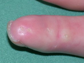
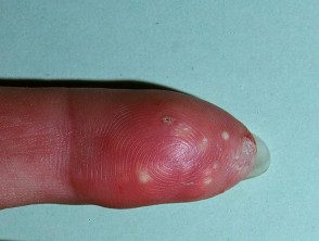
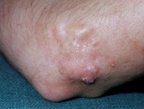
The signs and symptoms of calcinosis cutis vary depending on the underlying cause. In many cases, the lesions develop gradually and are often symptomless. Lesions usually appear as firm, whitish/yellowish papules, plates or nodules on the surface of the skin. A lonely injury can develop, although multiple lesions are more common. The lesions may become tender and ulcerate, discharging creamy, chalk-like material consisting primarily of calcium phosphate with a small amount of calcium carbonate.
Injuries to the tips of the fingers can be painful, while injuries elsewhere can restrict joint mobility and limit movement due to skin stiffness. In severe cases, cutaneous gangrene it can happen.
Calcinosis skin
Calcinosis skin
Calcinosis skin
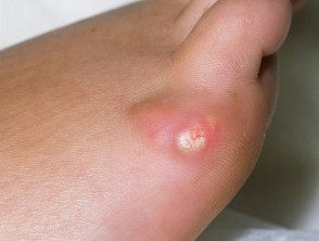
Calcinosis skin
How is calcinosis cutis diagnosed?
Laboratory tests are performed to determine any metabolic abnormalities that may lead to elevated calcium and phosphate levels. Radiological tests including plain x-rays, Connecticut Bone scan and scan are useful in demonstrating the extent of tissue calcification.
Biopsy of skin lesions is used to confirm the diagnosis. In histology of calcinosis cutis, granules and calcium deposits are seen in the dermis, often with a surrounding foreign body giant cell reaction. Calcium deposits can also be found in the subcutaneous tissue.
What is the treatment for calcinosis cutis?
The underlying cause of calcinosis cutis should be identified and treated accordingly. Medical therapy can be used to help relieve the symptoms of the condition, but they are generally of limited and variable benefit. Medications that may be tried include corticosteroids, probenecid, colchicine, sodium etidronate, diphosphonates, diltiazem, and magnesium and aluminum antacids.
Surgical removal of lesions is indicated when:
- It becomes very painful
- ulcerate and recurrent infections occur
- Cause functional impairment.
Because surgical trauma can stimulate further calcification, it may be best to remove a small site before proceeding with a large one. excision. Reappearance It is common after excision.
Depending on the underlying cause, a multidisciplinary team of doctors including nephrologist, rheumatologist, and hematologist may be necessary to manage the condition.
