

Ad
Skin cancer
Application to facilitate skin self-examination and early detection. read more.
What is it cutaneous scaly cell carcinoma?
Cutaneous squamous cell carcinoma (SCC) is a keratinocytes or notmelanoma skin Cancer formed by the uncontrolled growth and replication of squamous cells at epidermis. the curb produced by these cells is a corneal protein that normally forms the skin, hairand nail.
Cutaneous SCC is invader at dermis and rarely metastasis (spreads to other parts of the body). The most superficial forms of cutaneous SCC confined within the epidermis are known as intraepidermal SCC or SCC in the place.
Squamous cell carcinoma on white skin.
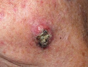
Squamous cell carcinoma
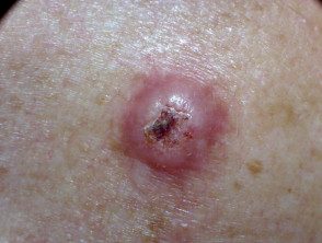
Squamous cell carcinoma
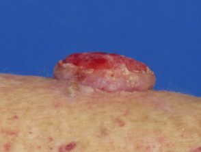
Squamous cell carcinoma
What is colored skin?
“Colored skin” is a subjective term that refers to natural skin that is “darker” than white (ie brown or black skin). When compared to a graded assessment of skin color types, such as Fitzpatrick phototypes, skin of color may refer to skin classified as type IV or higher [1]. In some contexts, colored skin is also used to describe the skin type of different non-white ethnic groups, including those of African, Asian, South American, Pacific Islander, Maori, Middle Eastern, and Hispanic descent. [2]. See ethnic dermatology.
Who gets cutaneous squamous cell carcinoma?
Cutaneous SCC is among the most frequently diagnosed cancers worldwide. [3].
-
Cutaneous SCC accounts for 15-25% of all diagnosed skin cancers and an estimated 75% of non-melanoma skin cancer-related deaths [3].
- The tallest incidence Cutaneous SCC is seen in fair-skinned individuals.
- Cutaneous SCC is the second most common form of skin cancer seen in Caucasians, Asians, and Hispanics after basal cell carcinoma
- It is the most common form of skin cancer seen in blacks and Asian Indians.
Incidence data
The incidence rates reported for cutaneous SCC vary according to the different studies and periods; Incidence rates per 100,000 population in the last 50 years have included:
- 0.16–2.9 in Chinese [4.5]
- 1.4 in Asian Indians [4]
- 3 in blacks (estimated) [6]
- 6 in New Zealand Maori (including basal cell carcinoma) [7].
Compared to skin of color, incidence rates of cutaneous SCC in fair-skinned people show a significantly stronger correlation with geographic location [8]. For example, Australia and New Zealand, which share a similar latitude, have the highest reported incidence of cutaneous SCC in the world, with reports of 228-1332 and 124-598 cases per 100,000 for Australia and New Zealand, respectively, in the middle. -1990 [3.8]. By comparison, incidence rates in Europe ranged from 9 to 28.9 per 100,000 in the population [3,8].
Overall incidence rates of cutaneous SCC are increasing, especially in fair-skinned individuals [8,9]. In recent decades, the incidence rate of cutaneous SCC has been estimated to have doubled or tripled in the United States, Australia, and Europe [9]. This trend is believed to be related to changes in popular recreational activities and clothing styles, resulting in an increase cumulative Sun exposure. The majority (~ 80%) of diagnoses occur in men and women over the age of 60, regardless of skin color. [8]. Men are much more likely than women to develop cutaneous SCC, with twice the incidence in some places. [8,9].
Risk factors for cutaneous SCC
Cutaneous SCC is more common in individuals with [10]:
- Previous SCC, basal cell carcinoma, or melanoma
- Actinic keratosis
- Outdoor occupations or recreational activities.
- A positive smoking state
- Fair skin, blue eyes, and blonde or red hair.
- Previous skin injury or scars from thermal burns or skin conditions (eg, cutaneous lupus erythematosus, epidermolysis bullosa, leg ulcers)
- Chronic ulceration
- Ionizing radiation, arsenic exposure, and immune deficiency due to disease (eg, Chronic lymphocytic leukemia) or immunosuppressive medications. Organ transplant Recipients are at massive risk of developing SCC.
Exposure to ultraviolet light (UV) Radiation B is a significant risk factor for cutaneous SCC in Caucasians; on skin of color, the scars may be more significant [3]. There is a 20 to 40% risk of cutaneous SCC in skin with chronic inflammation, ulceration or scarring leading to metastasis, compared with a 1-4% risk in sun-damaged skin [3].
What Causes Cutaneous Squamous Cell Carcinoma?
Cutaneous SCC is triggered by an abnormal disorder. genetic transformation.
More about mutations to DNA
In fair skin, oncogenetic transformation is related to sun exposure, with UVA and UVB radiation damaging and mutating the DNA of these cells. [9]. For example, cutaneous SCC cells have frequently shown mutations in p53 tumor suppressor gene - a gene that normally works to repair damaged DNA and prevent tumor development [8]. Mutations in this gene can alter its function, as p53 mutations are frequently found in many other types of malignant tumors. [8,9]. Similarly, UV exposure can move signaling pathways that regulate cellular activity, such as epidermal growth factor receiver (EGFR), RAS, Fyn or p16INK4a Pathways UV radiation can also suppress the immune system's ability to detect and destroy tumor cells. More significant immunosuppression, as in transplant patients, may allow infection by human papillomavirus strains implicated in causing cutaneous development of SCC [8].
In dark skin, a higher UV radiation absorption content. melanin believed to protect DNA from mutation and damages.
However, cutaneous SCCs can also arise in areas not exposed to sunlight or outside the skin. As the rates of these cutaneous SCCs may be higher in non-white skin ethical groups or show a hereditary pattern, it is believed that genetic processes outside the skin pigmentation influence the skin development of SCC [9]. For example, even in individuals with the same skin type, melanocortin receptor variants one (MC1R) the gene is believed to increase the risk of cutaneous SCC [9].
What are the clinical features of cutaneous squamous cell carcinoma?
Cutaneous SCC can occur anywhere on the skin. In people with fair skin, this usually occurs in areas frequently exposed to the sun. [3]. It is estimated that the 60-65% of cutaneous SCCs initially arise from actinic keratosis. Others may arise from intraepidermal carcinoma. In skin of color, cutaneous SCC most often develops in areas not exposed to the sun, such as the legs and feet, or on the anogenital region [3].
Cutaneous SCC:
- They are usually described as firm, elevated nodules or plates with a scab or scaly surface [3]
- Grow in weeks or months.
- It can be from millimeters to several centimeters in diameter.
- Can be cute
- It can also present as chronic non-healing ulcers [3].
Subtypes of Cutaneous Squamous Cell Carcinoma
Subtypes of cutaneous SCC include [10]:
-
Cutaneous horn, a conical projection of the skin formed by excessive keratin
-
Keratoacanthoma, rapid growth nodule with a central scale which can be reduced and resolved without treatment after a few weeks or months
- Carcinoma cuniculatum (warty carcinoma), a slow-growing, wart-like tumor on the sole of the foot
- Multiple SCC / keratoacanthoma-like eruptive lesions arising in syndromes (such as multiple self-healing squamous epitheliomas by Ferguson-Smith and Grzybowski syndrome)
Classification of cutaneous squamous cell carcinoma by risk
Cutaneous SCC can be classified as low or high risk; High-risk features of cutaneous SCC regardless of skin color include [10]:
- Emerging in greater o immunosuppressed individuals
- Location on the ear, lip, central face, hands, feet or genitals.
- Diameter ≥ 2 cm
- Histological thickness> 2 mm, bad differentiated histologyor with the invasion of subcutaneous tissue, nerves and blood vessels.
In colored skin, cutaneous SCC derived from perianal the scarred region or skin is at high risk due to relatively high metastasis rates [3].
More about risk
Doctors or pathologists can also define risk by the SCC subtype. Currently, there are no definitive classification systems used to classify cutaneous SCCs, although the following system has been proposed [11]:
- Low risk SCC:
- SCC arising from actinic keratosis
- SCC associated with HPV
- Trichilemmal carcinoma
- Spindle cell SCC not associated with radiation
- Intermediate risk SCC:
- Adenoid (acantholytic) SCC
- Intraepidermal epithelioma (Borst - Jadassohn) with invasion
- Lymphoepithelioma-like carcinoma
- High risk SCC:
- De novo SCC
- SCC arising in association with risk factors (radiation, burn scars, and immunosuppression)
- Invasive intraepidermal carcinoma (invasive SCC derived from intraepidermal carcinoma)
- Adenosquamous carcinoma
- Evil one proliferating trichilemmal cyst
- SCC of undetermined risk:
- Clear cell SCC
- SCC seal ring cell
- Papillary SCC
- Pigmented SCC
- Follicular SCC
- SCC derived from attached cysts
- Scaly eccrine ductal carcinoma.
How is cutaneous squamous cell carcinoma diagnosed?
Cutaneous SCC is usually suspected during a physical examination, often with the aid of a dermatoscope. In skin of color, particular attention may need to be paid to areas that are not normally exposed to the sun and to scarred or inflamed skin. [3].
Suspicious lesions are usually removed surgically to pathological examination and treatment (diagnosis excision)
Staging of cutaneous SCC
In patients with high-risk cutaneous SCC, it may be important to determine the depth of tumor invasion and spread to surrounding tissue (disease staging). The seventh edition of the American Joint Committee on Cancer (AJCC) staging system for cutaneous SCC is commonly used [12].
AJCC tumor staging for cutaneous SCC is as follows [12]:
- TX: Primary the tumor cannot be evaluated
- T0: No evidence of primary tumor
- Tis: Carcinoma in situ
- T1: Tumor ≤ 2 cm in greatest dimension with <2 características de alto riesgo
- T2: Tumor> 2 cm in greatest dimension with or without an additional high-risk feature, or any size with ≥ two high-risk features
- T3: Tumor with invasion of the maxilla, mandible, orbit or temporary bone
- T4: Tumor with invasion of the skeleton (axial or appendicular) or perineural invasion of the skull base.
(High-risk features include depth [> 2mmthickness; Clarklevel [> 2-mmthickness; Clarklevelofdermal invasion ≥ IV]; perineural invasion; location [primarydeloid site; primary site [primarysiteear; primarysitenon-glabrous lip]; Y differentiation [poorly differentiated or undifferentiated].)
The AJCC nodal The staging of cutaneous SCC is as follows [12]:
- NX: Regional lymph nodes cannot be evaluated
- N0: Not regional lymph node metastasis
- N1: Metastasis in a single ipsilateral lymph node, ≤ 3 cm in greatest dimension
- N2: Metastasis in a single ipsilateral lymph node,> 3 cm but not> 6 cm in greatest dimension; or in multiple ipsilateral lymph nodes, none> 6 cm in greatest dimension; or in bilateral or contralateral lymph nodes, none> 6 cm in greatest dimension
- N2a: Metastasis in a single ipsilateral lymph node,> 3 cm but not> 6 cm in greatest dimension
- N2b: Metastasis in multiple ipsilateral lymph nodes, none> 6 cm in greatest dimension
- N2c: Bilateral or contralateral lymph node metastases, none> 6 cm in greatest dimension
- N3: Lymph node metastasis,> 6 cm in greatest dimension.
(The sixth edition of the AJCC staging system classified regional lymph nodes in the absence [N0] or presence [N1] of lymph node metastases; the seventh edition incorporates another forecast factors, including the size, number, and location of the regional lymph node metastasis.)
To determine the stage of cutaneous SCC, the following investigations are generally considered in advanced cases:
- Ultrasound scans
- X-rays
- Connecticut scans
- Magnetic resonance scans
- Regional lymph node exams
- Lymph node biopsy
- A biopsy of other tissue.
Which is the differential diagnosis for cutaneous squamous cell carcinoma?
The main injuries that are confused with SCC are:
- Actinic keratosis
- Basal cell carcinoma
- Seborrheic keratosis
More information on differential diagnosis for cutaneous SCC
Other differential diagnoses for cutaneous SCC include:
Premalignant or malignant lesions:
- Actinic cheilitis (lip keratosis)
- Basosquamous carcinoma
- Melanoma
- Amelanotic melanoma
-
Oral intraepithelial carcinoma (oral lesions)
- Cutaneous T cell lymphoma
- Bowenoid Papulosis
- Atypical fibroxanthoma
-
Skin metastasis
Benign injuries:
- Seborrheic keratosis
- Common wart or wart
- Pyogenic granuloma
- Ecthyma
- Prurigo nodularis
- Trauma
- Thermal burn
Chronic conditions:
- Allergic contact dermatitis
- Atopic dermatitis
- Chronic lichen simplex
- Psoriasis
-
Lichen planus
-
Lupus erythematosus.
Malignant lesions, such as basal cell carcinoma and melanoma, have a peak incidence at the same approximate age (over 60 years) as cutaneous SCC, so they are more likely to be present simultaneously.
Cutaneous SCC can occur near or within the skin affected by chronic conditions (eg, psoriasis and lichen planus). In these cases, especially where the chronic condition is extended or poorly controlled, differentiating between injuries can be more difficult.
Compared with fair skin, cutaneous SCCs in skin of color show a more significant association with chronic disease. inflammatory or healing processes in the skin (Marjolin ulcer) [3].
What is the treatment for cutaneous squamous cell carcinoma?
Surgical excision is the recommended first-line treatment, regardless of skin color. [13]. Recommendations for cleavage margins are as follows [13]:
- Low risk, primary cutaneous SCC of less than <2 cm de diámetro con un margen de 4 mm de tejido circundante
- High risk, cutaneous SCC> 2 cm is removed with a margin of 6 mm of surrounding tissue, especially if they are:
- Classified as moderately or poorly differentiated
- Spreading into subcutaneous tissues
- Present on the ear, lip, scalp, eyelid, or nose
- Recurrent.
Other removal methods include [13]:
- Shaving, curettage and cauterizationand electrocautery for low-risk tumors in the trunk and extremities
- Cryotherapy for very small, thin, low-risk tumors
-
Mohs micrographic surgery for large facial lesions with confused margins or recurrent tumors
- Radiotherapy for inoperable tumors, patients unfit for surgery or as assistant to surgery.
Intraepidermal SCCs can be treated with current creams (fluorouracil or imiquimod) or cryotherapy if surgical removal is not adequate [13].
- Currently, there is insufficient evidence to determine whether cryotherapy or fluorouracil is a higher treatment [13].
- Please note that the use of imiquimod for intraepidermal SCC is off-license in New Zealand.
What is the treatment for advanced or metastatic squamous cell carcinoma?
Locally advanced primary, recurrent, or metastatic SCCs require multidisciplinary consultation. A combination of treatments can be used for these aggressive tumors, including:
- Surgery
- Radiotherapy
- Chemotherapy
- Experimental targeted therapy wearing epidermal growth factor receptor inhibitors (EGFRs) (these are not subsidized in New Zealand for use in cutaneous SCC).
The 2015 consensus-based European interdisciplinary guidelines for locally advanced or metastatic skin SCC make the following recommendations [14].
-
Surgery should include complete removal of the primary cutaneous SCC, in addition to in-transit or satellite metastases.
-
Radiation therapy should be administered, with or without chemotherapy, in cases where surgery is unsuccessful or infeasible. Radiation therapy can also be used as a palliative treatment to limit tumor growth. adjacent areas, relieve pain and stop any bleeding.
- There is no established chemotherapy regimen for cutaneous SCC. Stage IV (metastatic) cutaneous SCC has shown remission in response to chemotherapy; however, the evidence is limited and remission is often only seen for a short period of time.
- EGFR inhibitors can be considered after chemotherapy failure, but there is mixed evidence of their effectiveness.
Follow-up of cutaneous squamous cell carcinoma.
There are no established guidelines for follow-up. Follow-up is generally recommended since 30 to 50% of patients with a SCC are likely to develop another within five years. [14]. The time between follow-ups is generally based on the risk of new lesions, recurrences or metastasis. For patients with lower risk, annual follow-up may be sufficient. Patients at higher risk (eg, renal transplant patients with immunosuppressive drugs) may require closer follow-up [14].
Prevention of cutaneous squamous cell carcinoma in skin of color
Compared to fair-skinned people, the association between sun exposure and the development of cutaneous SCC is less prominent. In either case, a self-skin exam or skin checks performed by a physician can help identify skin SCCs earlier. Minimizing sun exposure is also potentially beneficial, since genetic processes outside of skin pigmentation can influence the risk of developing skin SCC [9]. Sun protection could involve:
- Staying indoors or in the shade in the middle of the day.
- Wear hedging clothing
- Generously applying broad spectrum topical sunscreens with protection factor SPF50 + to exposed skin yes outside
- Avoid tanning indoors (hammocks and solarium).
In individuals with multiple cutaneous SCCs or individuals at high risk of developing cutaneous SCCs, oral retinoids (ie, acitretin, isotretinoin) can delay tumor growth or the rate of development of new tumors [14]. However, these medications often exhibit side effects that, while not fatal, can be a nuisance in everyday life (i.e. dry skin and mucous membranes). They are also contraindicated in pregnancy due to a high risk of teratogenesis (congenital deformities).
What is the outcome for cutaneous squamous cell carcinoma of skin of color?
Successful treatment of cutaneous SCC is dependent about tumor size and invasion at the time of diagnosis. While most SCCs can be cured with treatment, people with skin of color tend to have more advanced cutaneous SCCs, especially if they occur on scarred or inflamed skin. As advanced cutaneous SCC are associated with lower survival rates, treatment outcomes for skin of color may be poorer [3].
More in forecast
Cutaneous SCCs> 20 mm in diameter or 2 mm in depth are associated with higher rates of reappearance or mortality. The 10-year survival rate in patients with cutaneous SCC that has spread to the lymph nodes has been estimated to be approximately 20% and <10% si se han producido metástasis a distancia. [3].
It is currently unknown why people with colored skin tend to have more advanced cutaneous SCCs compared to people with fair skin. This difference may be due to [3]:
- Late diagnosis
- Delayed presentation due to misperceptions related to skin cancer susceptibility
- A tendency for cutaneous SCC to be more aggressive in areas typically not exposed to the sun.
