
What is a dermatofibroma?
A dermatofibroma is common. benign fibrous nodule It most often arises on the skin of the lower legs.
A dermatofibroma is also called cutaneous fibrous histiocytoma
Who gets dermatofibroma?
Dermatofibromas occur at all ages and in people of all ethnicities. They are more common in women than in men.
What causes dermatofibroma?
It is not clear whether dermatofibroma is a reactive process or whether it is a neoplasm. The injuries are formed by proliferating fibroblasts. Histiocytes You may also be involved.
Sometimes they are attributed to an insect bite or a pink thorn injury, but not consistently. They may be more numerous in patients with impaired immunity.
What are the clinical characteristics of dermatofibroma?
Dermatofibromas most often occur on the legs and arms, but they can also arise on the trunk or anywhere on the body.
- People can have 1 or up to 15 injuries.
- The size varies from 0.5 to 1.5 cm in diameter; Most of the lesions have a diameter of 7 to 10 mm.
- They are firm nodules attached to the skin surface and movable over subcutaneous tissue.
- Skin dimples when pinching the injury.
- Color can be pink to light brown on white skin and dark brown to black on dark skin; some appear paler in the center.
- They usually do not cause symptoms, but are sometimes painful or itchy.
Dermatofibroma
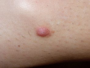
Dermatofibroma
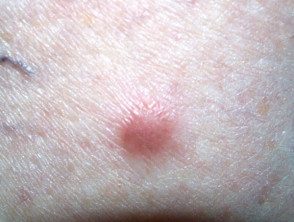
Dermatofibroma
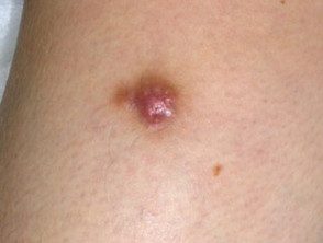
Dermatofibroma
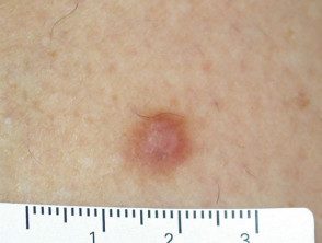
Dermatofibroma
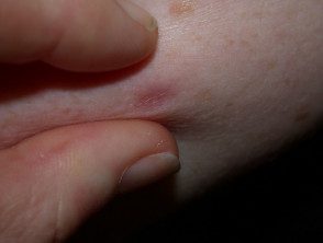
Pinch sign
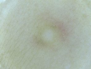
Dermoscopic view
See more images of dermatofibroma ...
Dermatofibroma complications.
Because they are often high injuries, they can be traumatized, for example, with a razor blade.
Occasionally, dozens can appear in a few months, usually in the context of immunosuppression (for example autoimmune disease, Cancer or certain medications).
Dermatofibroma does not lead to cancer. However, occasionally, it may be confused with dermatofibrosarcoma or desmoplastic melanoma.
How is dermatofibroma diagnosed?
Dermatofibroma is usually easy to diagnose clinically, with the support of dermoscopy. The most common dermoscopic pattern is a central white area surrounded by fainting. pigment net.
Diagnosis excision or skin biopsy takes place if there is a atypical feature like recent expansion, ulcerationor asymmetric structures and colors in dermoscopy.
the pathology of dermatofibroma shows swirls fascicles of spindle cell proliferation with excessive collagen statement at dermis. There are several pathological variants.
- cellular
- aneurysmal
- epithelioid
- atypical
- lipidized ankle type
- palisade
- cholesterotic
When in doubt, immunohistochemical staining is used to confirm the diagnosis.
What is the treatment for dermatofibroma?
A dermatofibroma is harmless and rarely causes symptoms. Usually only peace of mind is needed. If it is bothersome or cause for concern, the injury can be removed surgically.
Cryotherapy, shaving biopsy and To be Treatments are seldom completely successful.
