
What are angled lines?
In dermoscopy, angled lines are straight lines that meet at angles greater than 90 degrees but do not intersect. They can form total or partial polygonal Shapes are also known as polygons, rhomboids and zigzag patterns. They are a dermoscopic clue for melanoma.
What do angled lines look like through the dermatoscope?
Angled lines are gray-brown lines that are connected at an angle, or uniting to form polygons
angled lines
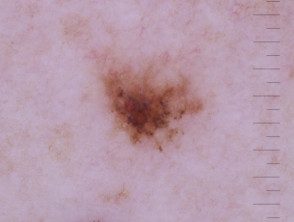
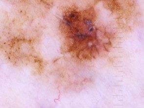
Angled lines seen on dermoscopy of a small melanoma
Angled lines are a clue to otherwise featureless melanoma
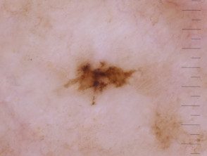
Angled lines seen on dermoscopy of a small melanoma
In which lesions are angled lines seen through the dermatoscope?
Angled lines are seen in the following skin lesions:
- Melanoma
- Lentigo evil
- Rarely, pigmented basal cell carcinoma.
On the face, angled lines correspond to rhomboids and are also represented in:
- Pigmented actinic keratosis
- Lichen planus like keratosis.
Dermoscopic angular lines
Angled lines seen in dermoscopy of a melanoma
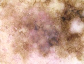
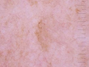
Angled lines seen on dermoscopy of lentigo maligna
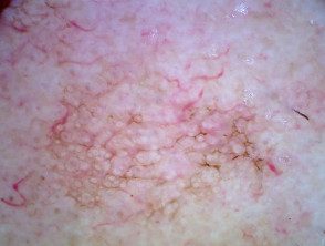
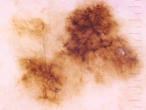
Angled lines observed in dermoscopy of actinic keratosis.
Angled lines seen on melanoma dermoscopy
Which is the histological explanation of angled lines?
The precise histology is unclear, but probably correlates with an accumulation of inflammatory cells and melanophages at dermis, under the evil one melanocytes that resides in the epidermis.
The angled lines seem to correspond to a flattened dermoepidermal junction (DEJ) with less and more blunt challenge pins due to proliferation of atypical melanocytes in the DEJ, as well as a focal accumulation of melanophages in the superficial dermis [1].
