
What is favus?
Favus or tinea favosa is in most cases a severe form of tinea capitis. It is caused by the dermatophyte fungus Trichophyton schoenleinii and results in the destruction of the honeycomb. hair axis. Although rare, it can sometimes occur as onychomycosis, tinea barbae, or tinea corporis.
No cases of favus have been reported in New Zealand. It is also relatively rare in the United States, and most cases are limited to areas where malnutrition, poverty, and poor living conditions exist. Favus has been found in Poland, southern and northern Africa, Australia, Pakistan, Brazil, the Middle East, and the United Kingdom.
Favus
Courtesy of Dr. Mohammad Mahmoud.
Who does favus affect?
Favus affects both men and women and occurs in both children and adults. Often the infection it is trapped during childhood or adolescence and persists into adulthood. It does not appear to be highly contagious, but it is not uncommon for various family members to become infected, particularly where people live in close proximity to one another.
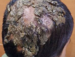
How does favus look?
Favus is characterized by yellow cup-shaped crusts (scutula) that are grouped in patches like a piece of honeycomb, hence the name favus (from the Latin “honeycomb”). Every Cortex it surrounds a lock of hair, which pierces the center and protrudes.
There appear to be 3 stages of the disease depending on its severity.
- Stage 1: area around the hair. follicles on the scalp it becomes red and inflamed. The hair remains intact.
- Stage 2: Yellow cup-shaped crusts (scutula) form and hair begins to fall out.
- Stage 3: at least one third of the scalp is affected and there is a great deal of hair loss, atrophy and scars
The shape of the dome is dense. plates they often host secondary bacterial infection. Removal of the plates leaves a wet, red and swollen base. There is often a mouse smell.
Some other forms of favus related to their clinical characteristics have been described:
- Favus pityroides: mimics dandruff or seborrheic dermatitis
- Favus psoriasiformis: favus that mimics psoriasis
- Follicular Favus: Cone-Shaped Wax Color papules around the follicles
- Favus impetigoides: yellowish crusts that mimic impetigo lesions
- Favus papyroides - small loci on the scalp covered by a brittle substance
- Favus herpetiformis: round, erythematous, climbing plaques with small papules, vesicles, pustules and crusts found on the trunk and extremities.
Diagnosis
The diagnosis of favus must be confirmed by microscopy and culture skin scrapings and hair plucked from the roots (see laboratory tests).
What is the favus treatment?
Favus requires treatment with an oral antifungal agent. As used in other types of tinea capitis, griseofulvin is probably the most effective agent for infection with Trichophyton schoenleinii, but it is no longer available in New Zealand. More recent clinical studies indicate that Trichophyton schoenleinii infections can be successfully eradicated using oral terbinafine, itraconazole, or fluconazole. Treatment with these antifungal agents may require a longer course of treatment for ringworm of the head.
In addition to systemic treatment, current Agents such as ketoconazole 2% and selenium sulfide 2.5% shampoo may be useful. Debris and scabs from the scalp should be removed and the scalp should be cleaned regularly.
Favus is essentially a chronic disease and can last from 10 to 20 years. If not properly diagnosed and treated, it can lead to alopecia (hair loss) and extensive scarring.
