
What is it granulomatous dermatitis?
Granulomatous dermatitis describes several disorders characterized by their histological appearance.
- Interstitial granulomatous dermatitis (IGD)
-
Palisading neutrophilic granulomatous dermatitis (PNGD)
-
Interstitial granulomatous pharmacological reaction (IGDR)
Interstitial granulomatous dermatitis
Interstitial granulomatous dermatitis is a rare skin disorder in which there is a particular pattern of granulomatous. inflammation.
The classic original clinical description of interstitial granulomatous dermatitis was linear erythematous palpable laces about him side aspects of the trunk, called 'the rope sign'. However, several different types of eruption Since then they have been described with the same histological aspect.
What are the clinical features of interstitial granulomatous dermatitis?
The characteristics of interstitial granulomatous dermatitis are variable.
- Red or skin-colored patches papules and plates
- The shape of the lesions can be round, cancel or cable-I like it.
- Lesions increase and decrease, and can vary in size and shape over days or months.
- They usually have no symptoms, but some patients complain of mild itching or burning sensation.
- Lesions tend to be symmetrically distributed on the trunk, but proximal the extremities may also be affected.
- It most often affects middle-aged women.
- Many affected patients also suffer from autoimmune disease.
Interstitial granulomatous dermatitis
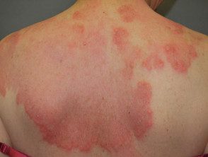
Interstitial granulomatous dermatitis
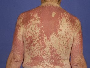
Interstitial granulomatous dermatitis
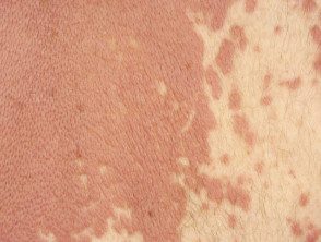
Interstitial granulomatous dermatitis
How is interstitial granulomatous dermatitis diagnosed?
Interstitial granulomatous dermatitis is diagnosed by a pathologist when examining a skin biopsy. The characteristic histological features of interstitial granulomatous dermatitis are:
- Dense histiocytic inflammation in the lattice (lower) dermis
- Scarce neutrophils and eosinophils
- Perivascular and interstitial lymphocytes
- Interstitial histiocytes (Between the collagen bundles) or palisated histiocytes (aligned perpendicular to the fragmented central collagen).
- Focal Collagen degeneration, which may be surrounded by space (floating sign)
- Few giant cells.
How does interstitial granulomatous dermatitis compare to granuloma annulare?
Granuloma annulare and interstitial granulomatous dermatitis may appear clinically similar and histologically. Granuloma annulare also presents with papules and plaques, but they are usually found on the back of the hands or feet, while the trunk is a more common site for interstitial granulomatous dermatitis. Granuloma annulare is less frequently associated with autoimmune diseases.
The characteristic histological features of ring granuloma are:
- Histiocytes located in the upper dermis
- Rare or absent neutrophils and eosinophils
- Abundant mucin [1].
Pale neutrophilic granulomatous dermatitis
Paletrating neutrophilic granulomatous dermatitis was first described as scab umbilical papules on the elbows that arise in patients with rheumatoids arthritis (Winkelmann's granuloma) and eosinophilic granulomatosis with polyangiitis (Churg-Strauss syndrome)
Several different types of rash with the same histologic appearance have been described, including annular plaques on the trunk. Lesions are often tender and can ulcerate.
How is palisade neutrophilic granulomatous dermatitis diagnosed?
Pale neutrophilic granulomatous dermatitis is diagnosed by skin biopsy. The characteristic histological features of palisade neutrophilic granulomatous dermatitis are:
- Intense neutrophils infiltrate and nuclear dust
- Interstitial histiocytic infiltrate
- Collagen degeneration
- Leukocytoclastic vasculitis
- Variable histological aspect according to the stage of the eruption.
Palisading granulomas Seen they have also been described as miniature “Churg-Strauss granulomas” or llama figures, with degenerate collagen enveloped by eosinophils that resemble a llama. Flame figures are also seen on the pathology Wells syndrome.
What are the clinical associations of granulomatous dermatitis?
Several conditions associated with interstitial granulomatous dermatitis have been observed to arise. There is less information on associations with palisade neutrophilic granulomatous dermatitis.
Autoimmune diseases and conditions
Both forms of granulomatous dermatitis have often arisen in people with other conditions considered to be of autoimmune origin, implying that an immune complex mechanism may be involved in the Pathogenesis [two]. These have included the following conditions.
-
Rheumatoid and non-rheumatoid arthritis, characteristically symmetrical and involving the fingers, wrists, elbows and shoulders, it is the most common association. Arthritis (swollen joints) or arthralgia (painful joints) can occur before, during or years after the onset of skin lesions in approximately 50% of published cases of interstitial granulomatous dermatitis [3].
- the systemic lupus erythematosus (SLE)
- Primary antiphospholipid syndrome
- Churg-Strauss syndrome
- Thyroiditis
- Vitiligo
In a literature review of 15 patients with interstitial granulomatous dermatitis, autoantibodies identified in blood tests included rheumatoid factor (RhF), antinuclear factor (ANA), thyroglobulin, histone SS-A, DNA histone and HAUNCH [4].
Malignancy
There are some reports of association of malignancy with interstitial granulomatous dermatitis. In one case, the lesions disappeared after the lung. Cancer was treated [5]. Rare associations of granulomatous dermatitis with leukemia [6], lymphoma, breast cancer, hypopharyngeal scaly cell carcinoma and endometrial neoplasm Has been reported.
How is granulomatous dermatitis treated?
Granulomatous dermatitis usually becomes inflamed and remits. Successful treatments have included:
- Current steroids
- Hydroxychloroquine
-
Dapsone
One case associated with SLE responded well to systemic steroids after 15 days. [two].
Although tumor necrosis factor (Alpha TNF inhibitors) have recently been described as inducers of interstitial granulomatous dermatitis [7], Etanercept has been used in interstitial granulomatous dermatitis associated with rheumatoid arthritis with complete skin clearance and improvement in arthritis [8].
Interstitial granulomatous pharmacological reaction
Drug-induced interstitial granulomatous dermatitis is known as an interstitial granulomatous drug reaction. It is believed to be a distinct clinical and pathological entity. It occurs as annular plates, and nodules in the trunk, arms, medium thighs and skin folds. The rash resolves when the responsible medication is withdrawn.
How is an interstitial granulomatous drug reaction diagnosed?
The interstitial granulomatous drug reaction is diagnosed by skin biopsy. The characteristic histological features of the interstitial granulomatous drug reaction are:
- Basal epidermal cell vacuolization
- Lichenoid changes with eosinophils
- Absence of neutrophils.
- In some cases, palisade granulomatous changes associated with collagen necrosis and floating sign.
Medications that have been reported to be involved in the eruption of interstitial granulomatous drugs include:
-
TNF-alpha inhibitors (infliximab, adalimumab, etanercept) [7]
- Beta blockers
- Calcium channel blockers
- Angiotensin conversion enzyme inhibitors
- Lipidflowering agents
- Statins (HMG-CoA reductase inhibitors)
- Frusemide
- Antidepressants
-
Anticonvulsants [4].
