
What is leishmaniasis?
Leishmaniasis is a parasitic disease transmitted by sand flies infected with protozoa Leishmania. Leishmaniasis is endemic in more than 70 countries worldwide and affects about 12 million people.
There are several clinical forms of leishmaniasis. The clinical manifestation of the infection depends on the species Leishmania, which varies with the geographic area and the hostThe immune response.
The parasites that cause human leishmaniasis are not found in New Zealand, Australia, the South Pacific, or Antarctica.
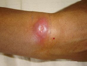
old world leishmaniasis
See more images of leishmaniasis.
Who gets leishmaniasis?
People of all ages who live in or travel through areas where sandflies and Leishmania species are endemic are at risk for infection with leishmaniasis. Living in rural areas and spending time on or near the ground increases your risk.
Classification and causes of leishmaniasis.
There are more than 20 species of Leishmania parasites that can infect humans; transmitted through the bite of phlebotomine. Sand flies are small (1.5–3 mm) insects that actively feed on blood at dawn and dusk. Sand flies live in wall crevices, animal burrows, and leaf litter, in tropical and subtropical regions. his bite is asymptomatic and classically in exposed sites.
Leishmaniasis has several recognized clinical forms, and its manifestation depends on the species. inoculated and the host immune response. The most important distinction is between American and non-American species of Leishmania, As the viannie subspecies found in the Americas, may result in mucocutaneous leishmaniasis
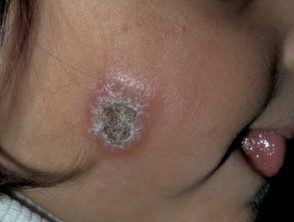
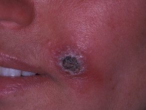
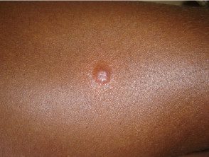
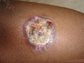
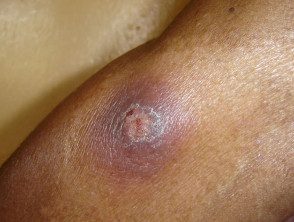
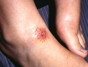
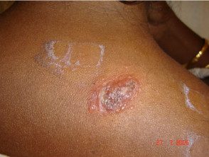
Cutaneous leishmaniasis
Cutaneous leishmaniasis usually occurs at the site of inoculation. the presentation and forecast will vary depending on the species involved.
Non-American (old world) cutaneous leishmaniasis:
- Middle East, North Africa, Asia
- L major, L tropica, L infantum, L donovani
- Synonyms: Oriental pain, Baghdad boil, Delhi boil, Saldana, Aleppo button, granuloma endemic
American (New World) cutaneous leishmaniasis:
- Central and South America
- L mexicana, L braziliensis, L amazonensis
- Synonyms: chiclero ulcer, uta, Bauru ulcer, pian boi, liana.
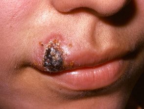
Mucocutaneous leishmaniasis
Mucocutaneous leishmaniasis is a destructive form of leishmaniasis, seen only with the American species of Leishmania (viannie subspecies).
American mucocutaneous leishmaniasis:
- Central and South America
- L. braziliensis, L. guyanensis, L. panamensis
- Synonym: espunda.
Diffuse cutaneous leishmaniasis
Diffuse cutaneous leishmaniasis is a rare presentation that results from an anergic host response to the parasite.
Non-US diffuse cutaneous leishmaniasis:
- Ethiopia, Kenya
- L aethiopica.
American diffuse cutaneous leishmaniasis:
- South America
- L. amazonensis.
Visceral leishmaniasis
Visceral leishmaniasis is the result of internal organ involvement and is generally fatal if left untreated. It is also known as kala-azar or dumdum fever.
Non-American visceral leishmaniasis:
- India, Southern Europe, China, North Africa, Kenya
- L donovani, L infantum, L tropica.
American visceral leishmaniasis:
- Central and South America
- l chagasi.
Post-kala-azar dermal leishmaniasis
Post-kala-azar dermal leishmaniasis is a form of cutaneous leishmaniasis that can occur months to years after treatment for visceral leishmaniasis.
Non-US post-kala-azar dermal leishmaniasis:
- sudan, india
- L donovani, L infantum, L tropica.
American post-kala-azar dermal leishmaniasis:
- Central and South America
- L chagasi.
relapsing leishmaniasis
Relapsing leishmaniasis is a rare, cutaneous form of leishmaniasis, which occurs in patients with good cellular immune response. It is also known as lupoid leishmaniasis.
Non-US relapsing leishmaniasis:
- Middle East, India, Southern Europe
- He tropics.
American relapsing leishmaniasis:
- Central and South America
- L. braziliensis.
What are the clinical features of leishmaniasis?
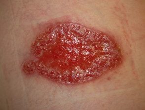
Cutaneous leishmaniasis
- Cutaneous leishmaniasis is the most common form of leishmaniasis.
- Solitary lesions are typical, but multiple lesions occur
- the initial injury it's a small red papule, gradually enlarging to 2 cm in diameter
- Central ulceration it is typical
- Ulcers may be moist and ooze pus or dry with a crusty crust
- The sores usually appear on exposed areas of the skin, especially on the face and extremities.
- the incubation the time between an infected sand fly bite and the development of the lesion is typically 2 weeks to 6 months
- The lesions are generally painless and most resolve spontaneously, often leaving residue. atrophic scars
- The resolution time varies between 2 months and more than a year.
- Sporotrichoid spread with lymphocutaneous nodules can happen
- Chronic disease can occur and there is a risk of dissemination in immunocompromised patients
Mucocutaneous leishmaniasis
- Mucocutaneous leishmaniasis typically occurs after spontaneous resolution or local treatment of the leishmaniasis. primary skin lesion
- It can develop in months or after many years.
- The lifetime risk of developing mucocutaneous leishmaniasis after skin injury caused by L. braziliensis it is around 5%
- The lesions typically affect the mucous membranes of the nose and mouth, but the mucous membrane the surfaces of the eyes and the genital tract may also be involved
- Without treatment, ulceration of the mucosal surfaces and destruction of the underlying tissue may occur.
- Mucosal lesions are often painful and become sites of infection, sometimes leading to septicemia
- It can cause extreme disfigurement.
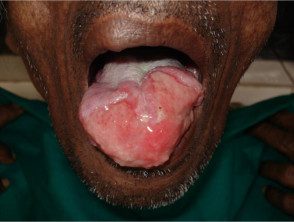
mucosal leishmaniasis
Diffuse cutaneous leishmaniasis
- Diffuse cutaneous leishmaniasis is a specific disease entity; sometimes the term is used incorrectly to describe disseminated or multiple cutaneous leishmaniasis
- Results of an anergic response to infection due to reduced cellular immunity
- After primary cutaneous leishmaniasis lesion, non-ulcerative nodules and plates develop
- Lesions can be numerous and spread throughout the body
- Follow a chronic relapse or progressive grade
- Often difficult to treat
Visceral leishmaniasis
- Visceral leishmaniasis affects the internal organs, including the spleen, liver, and lymph nodes
- Signs and symptoms include fever, weight loss, lymphadenopathy, hepatomegaly and massive splenomegaly
- Laboratory tests may show pancytopenia and hypergammaglobulinemia
- Complications include gastrointestinal bleeding, peripheral edema, acute renal failure and secondary bacterial infections
- Generalized hyperpigmentation it is a late feature of visceral leishmaniasis; the other name for it, kala-azar, comes from the Hindi for "black fever."
- Visceral leishmaniasis can take different forms, from a self-resolving asymptomatic disease to a fulminant and life-threatening disease.
Post-kala-azar dermal leishmaniasis
- Post-kala-azar is a cutaneous form of leishmaniasis that results as a complication of visceral leishmaniasis.
- It occurs months or years after treatment for visceral leishmaniasis.
- Most cases occur in Sudan (up to 50%) and India (up to 10%)
- injuries are indurated papules and nodules or areas of macular hypopigmentation
- It most often affects the face, trunk, and extremities.
- Oral mucous membrane and the genital area may also be involved
- Prognosis and treatment vary by location: in more than 80% of Sudanese patients, post-kala-azar dermal leishmaniasis lesions will resolve spontaneously within a year, while in India the rate of spontaneous resolution is much lower , so that systemic therapy is used before
relapsing leishmaniasis
- Relapsing leishmaniasis occurs in patients with a good cellular immune response.
- Spontaneous resolution of the primary skin lesion is followed by the development of new lesions around the edge of the primary lesion. scar
- Lesions typically ulcerate and then heal
- The cycle continues with a chronicle recurrent of course, usually for decades
Cutaneous leishmaniasis in Sri Lanka
How is cutaneous leishmaniasis diagnosed?
The diagnosis of cutaneous leishmaniasis is generally based on the history and clinical appearance of the lesion. A complete travel history, including historical travel due to the long incubation period, is important in non-endemic areas. The diagnosis can be confirmed by identifying the parasite in biopsy or division of skin smears. Culture and PCR It can also be used to confirm the diagnosis and identify Leishmania species, which is important when there is a risk of mucocutaneous leishmaniasis.
Serology It is used to confirm the diagnosis in cases of visceral leishmaniasis.
In more than 70% of cases, a full-thickness skin biopsy can reveal the parasite. Histopathology It is also used to establish mucocutaneous leishmaniasis and visceral leishmaniasis. Complete blood counts and liver function tests should also be done in visceral leishmaniasis.
New World leishmaniasis
Sore
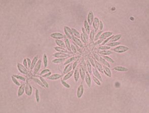
Lieshmania viannia promastigotes
Sore
Which is the differential diagnosis for leishmaniasis?
The variety of clinical manifestations of cutaneous leishmaniasis results in a wide range of differential diagnoses:
- Insect bites
- Nodular chondrodermatitis helicis
- Basal cell carcinoma
- Scaly cell carcinoma
- Granuloma annulare
- Atypical mycobacterial infection
- Vulgar lupus
- Leprosy
- Actinomycosis
- Tularemia
- Melioidosis
- deep fungal infections
- Sporotrichosis
- cutaneous anthrax
- gangrenous ecthyma.
What is the treatment for leishmaniasis?
In cutaneous leishmaniasis, treatment options differ depending on whether the lesion/s is considered simple or complex.
In non-US cutaneous leishmaniasis, lesions are considered complex if they are larger than 4 cm, multiple, associated with lymphatic spreading, persistent (> 6 months), in immunosuppressed individuals, or located over joints or in cosmetically sensitive areas.
All cases of US leishmaniasis should be considered complex due to the risk of mucocutaneous leishmaniasis with the viannie subspecies.
Treatment options for cutaneous leishmaniasis lesions include:
- Self-heal (simple injuries only)
-
Current non-antimonal treatments
- Cryotherapy
- heat therapy
- Photodynamic therapy
- Imiquimod
- Topical paromomycin (also known as aminosidine)
- intralesional antimonials
- sodium stibogluconate
- meglumine antimony
- Non-antimonal systemic therapies
- Amphotericin B
- Miltefosine
- Pentamidine
- Azole antifungal drugs: itraconazole, fluconazole, ketoconazole
- Paromomycin
- Zinc sulfate
- Alopurinol
- Systemic antimonials (intravenous or intramuscular)
- sodium stibogluconate
- Meglumine antimony.
Most cases of simple cutaneous leishmaniasis will resolve on their own without treatment, but this can take many months and can lead to scarring.
Systemic antimonials are the mainstay of treatment for complex lesions of cutaneous leishmaniasis, mucocutaneous leishmaniasis, and visceral leishmaniasis. They cannot be administered orally, and the duration of treatment can be up to 28 days for mucosal lesions. Treatment requires hospital admission and there is a risk of side effects, including cardiotoxicity.
Secondary wound infections should be treated with antimicrobial therapy.
Surgical treatments and vaporization To be Treatments are also used for appropriate injuries.
Consult the guidelines on the treatment of leishmaniasis published by the Infectious Diseases Society of America (IDSA) and the American Society of Tropical Medicine and Hygiene (ASTMH) (December 2016).
Prevention of insect bites.
Infection can be prevented by avoiding sand fly bites. Because there are currently no vaccines or drugs to prevent infection, travelers to areas where leishmaniasis is present predominant You should decrease your risk of being bitten by taking the following precautionary measures.
- Avoid outdoor activities, especially from dusk to dawn when sand flies are most active.
- Wear long-sleeved shirts, long pants, and socks: tuck shirt into pants.
- Apply insect repellent on exposed skin and under the ends of the sleeves and the legs. The most effective repellents are those that contain the chemical DEET (N,N-diethyl-meta toluamide).
- Spray Clothing, living and sleeping areas (including bed netting) with insecticides containing permethrin.
