

Ad
Skin cancer
Application to facilitate skin self-examination and early detection. read more.
What is it amelanotic melanoma?
Amelanotic melanoma is a form of melanoma in which the evil one cells have little or nothing pigment. The term 'amelanotic' is often used to indicate lesions that are only partially devoid of pigment, whereas truly amelanotic melanoma where lesions are devoid of all pigment is rare [1].
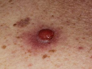
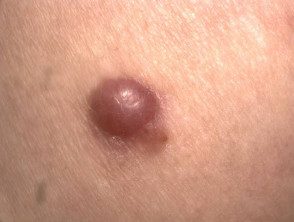
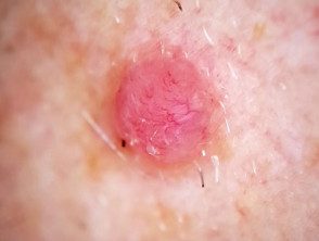
Amelanotic melanoma
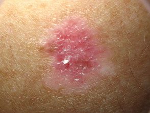
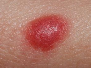
Nodular melanoma
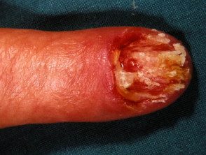
Ungual melanoma
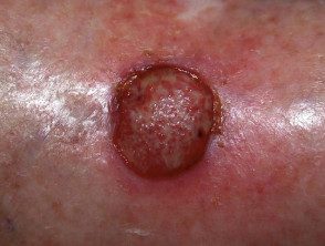
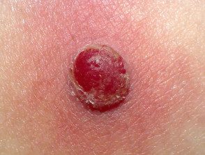
Ulcerated melanoma
See more images of amelanotic melanoma
Who gets amelanotic melanoma?
Amelanotic melanoma accounts for approximately 1–8% of all melanomas. the incidence of truly amelanotic melanoma is difficult to estimate, since many hypopigmented lesions are labeled amelanotic [1].
Risk factors for developing amelanotic melanoma include:
- Increasing age, although amelanotic melanoma accounts for a significant proportion of melanoma in young children [2]
- Sun-exposed skin - particularly in older people with chronic Photodamage.
What Causes Amelanotic Melanoma?
Melanoma is caused by malignancy melanocytes. The development of malignancy in melanocytes it is due to genetic changes in DNA, but how and why this occurs is largely unknown. The melanoma cells in amelanotic melanoma cannot produce mature ones. melanin granules, which results in lesions lacking pigment.
To explain the lack of pigment, three models have been proposed.
- Amelanotic melanoma can be a bad differentiated typical melanoma subtype.
- Amelanotic melanoma can be a dedifferentiated melanoma that has lost its normality. phenotype.
- Amelanotic melanoma cells can retain their melanocytic identity but gain the ability to form different phenotypes (multipotency) [3].
What are the clinical features of amelanotic melanoma?
Amelanotic melanomas are classically described as 'skin-colored'. A significant proportion is red, pink or erythematous. Typical early lesions present as asymmetric macular lesions that may be uniformly pink or red and may have a slight light tan, brown, or gray pigmentation on the periphery Borders can be well or poorly defined [4].
Any subtype of melanoma can be amelanotic.
- Nodular melanoma may look like a pink or rosy melanocytic nevus (mole) or basal cell carcinoma.
- With subungic melanoma, 25% are amelanotic
- With desmoplastic melanoma, more than 50% are amelanotic [5].
- Metastatic melanoma does not present uncommonly with amelanotic lesions even when the primary injury it is pigmented [5].
Amelanotic melanomas may not show the ABCDE clinical criteria (asymmetry, border irregularity, color variation, large diameter) that are classically used as warning signs of melanoma. Patients and clinicians may not be alert to suspect non-pigmented lesions such as melanoma, and therefore amelanotic melanomas are often misdiagnosed. Expanding the ABCD warning signs to include the 3 Rs (red, elevated, recent change) may help detect amelanotic melanoma [6].
How is amelanotic melanoma diagnosed?
Clinical features
Amelanotic melanomas have little or no pigment and can be clinically difficult to diagnose.
A patient's history of observing a change in a lesion is an important diagnostic factor with amelanotic lesions, and amelanotic melanoma should be considered in the differential diagnosis.
Examination of the entire skin surface is also important, since sun damage (eg, actinic keratosis) and other pigmented lesions may provide clinical clues that an unpigmented or hypopigmented lesion may be an amelanotic melanoma.
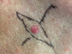
Clinical characteristics of melanotic melanoma.
Superficial spreading amelanotic melanoma in situ
Amelanotic nodular melanoma
Amelanotic nodular melanoma
Dermoscopy
Dermoscopy, the examination of a lesion using a dermatoscope, can be helpful in diagnosing suspicious lesions. Vessel morphology it must be carefully evaluated if the lesions lack pigmentation.
The characteristic dermoscopic features of amelanotic melanoma are:
- Irregular pigmentation (if any present)
- Irregular dots or blood cells (in pigmented areas)
- Polymorphous vascular patterns including milky red areas, lattice depigmentationdotted, irregular vessels, linear irregular vessels, or the combination of irregular dotted and linear vessels (the most common dermoscopic finding)
- white lines [1].
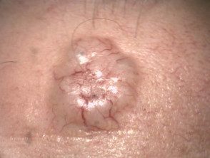
Melanoma dermoscopy (same lesions as before)
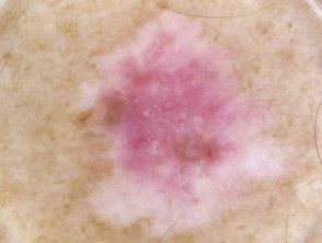
Superficial spreading melanoma in situ
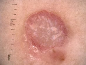
Nodular melanoma
Nodular melanoma
Biopsy
Lesions suspected of amelanotic melanoma should be excised with a clinical margin of 2–3 mm and sent for pathological diagnosis (excision biopsy).
Amelanotic melanoma may have varied histopathological appearances. Amelanotic melanoma can be masked as a series of non-melanocytic neoplasms [7].
- A key feature is the lack of melanin granules; spots that detect melanin granules include the Fontana-Masson stain [3].
-
Immunohistochemical staining can reveal some residual pigment in amelanotic melanoma and identify malignant cells as melanocytes.
- Electron microscopy can be used to demonstratemelanosomes in unclear cases
Pathology report
the pathologistThe report presents both a macroscopic and microscopic Description of the excised lesion and includes:
- Diagnosis of primary melanoma
- Breslow thickness to the nearest 0.1 mm
- Clark's invasion level
- Excision margins (normal tissue around a tumor)
- the mitotic rate (how fast are cells proliferating)
- Whether or not there is ulceration.
The report may also include comments on the cell type and its growth pattern, invasion of blood vessels or nerves, inflammatory reply, regression, immunohistochemistryand if there is an original associated benign melanocytic lesion [8].
What is the thickness of Breslow?
The Breslow thickness is reported for invader melanomas It is the vertical measurement in millimeters from the top of the granular layer (or base of superficial ulceration) to the deepest point of tumor involvement. The thickness of Breslow is the most important local forecast factor in primary melanoma, as thicker melanomas are more likely to metastasis (to spread) [8,9].
What is Clark's level of invasion?
The Clark level indicates the anatomical plane of the invasion. This is useful for predicting the outcome for thin tumors and less useful for thick ones compared to the Breslow thickness value. [[8,9 9].
What is the differential diagnosis for amelanotic melanoma?
Clinically, amelanotic melanoma may present as an erythematous scaly taint, license plateor nodule with irregular edges, mimicking numerous other skin lesions such as:
- Basal cell carcinoma
- Spitz naevus
- Seborrheic keratosis
- Actinic keratosis
- Pyogenic granuloma
- Dermatofibroma
- Keratoacanthoma
- Intraepidermal scaly cellular carcinoma [1,4].
The differential diagnosis of amelanotic melanoma.
Noducytic basal cell carcinoma
Spitz naevus
Pyogenic granuloma
What is the treatment for amelanotic melanoma?
Amelanotic melanoma is treated in the same way that a pigmented melanoma is treated.
After diagnostic excision, the next step is wide local excision of the wound with a 10-20 mm margin of normal tissue. The extent of the surgery depends on the Breslow thickness of the melanoma and its site. Amelanotic melanomas can be incompletely excised despite the recommended margins, as the margins are often difficult to define. An understanding histological The exam that includes immunohistochemical staining helps determine the edge of a tumor. A new split may be necessary. Recommended margins in provisional 2013 New Zealand Melanoma Service Delivery Standards are as follows [10]:
-
Melanoma in the place (Tis): 5–10 mm
- Melanoma <1 mm (T1): 10 mm
- Melanoma 1–2 mm (T2): 10–20 mm
- Melanoma 2–4 mm (T3): 20 mm
- Melanoma> 4 mm (T4): 20 mm [9]
Sentinel lymph node The biopsy can be discussed with patients with melanomas thicker than 0.8 mm. This can provide staging and prognostic information [9].
Amelanotic melanoma can metastasize (spread to distant sites such as lymph nodes or in another part of the body). These cases require individualized treatment that may include surgery, radiotherapy, chemotherapyor targeted therapy.
What happens in the follow-up of amelanotic melanoma?
Follow-up is an important part of the treatment of amelanotic melanoma because it allows the opportunity to detect recurrences early. It also allows the opportunity to detect new primary melanomas as early as possible. A second invasive melanoma occurs in 5-10% of patients, and an unrelated melanoma in situ affects more than 20% of patients [8].
2008 Clinical Practice Guidelines for the Management of Melanoma in Australia and New Zealand We recommend the following for the follow-up of patients with invasive melanoma:
-
Self skin exams are performed.
- Periodic skin checks should be done with the patient's preferred healthcare professional.
- Follow-up intervals are preferably 6 months for 5 years for patients with stage 1 disease, 3 months or 4 months for 5 years for patients with stage 2 or 3 disease, and annually thereafter for all patients.
- The individual needs of the patient should be considered before offering adequate follow-up.
- Education and support should be provided to help the patient adjust to their illness. [[8,9 9].
What is the outcome of amelanotic melanoma?
the forecast of amelanotic melanoma is similar to that of pigmented melanomas. Prognostic factors include the Breslow thickness of the melanoma at the time of excision (this is the most important factor), the location of the lesion, the patient's age, and sex. It is important to note that due to its atypical clinical features, amelanotic melanomas may have a delay in their diagnosis and are consequently often more advanced than pigmented melanomas when diagnosed [5].
The risk of metastasis it is directly related to Breslow's thickness, with thicker melanomas more likely to metastasize. 2008 Clinical Practice Guidelines for the Management of Melanoma in Australia and New Zealand report that metastasis are rare for thin melanomas (<0,75 mm), con un riesgo que aumenta al 5% para melanomas de 0,75–1,00 mm de espesor. Los melanomas más gruesos de 4.0 mm tienen un riesgo significativamente mayor de metástasis del 40% [9].
