Category

Aesthetic medicine blog

How light damages and the importance of protecting yourself from it

porphyrias-protect-light-9536448-9678870-jpg-2362796

Knowledge of Photodynamic Therapy helps to understand how light damages people with porphyrias and the importance of protecting themselves from it.

One afternoon, while I was in consultation, a young patient came to see me with his father. The patient had a porphyria, a strange disease, but after chatting with them, I realized that my years of dedication to photodynamic therapy could help these patients. And this is the reason why I write this text on my blog. I hope you find it useful.

Porphyrias are rare diseases and are characterized by a exaggerated photosensitivity (sun damage).

Dermatologists have “copied” this mechanism in Photodynamic therapy (PDT) to produce phototoxicity located in tumor and momentary lesions, which, by applying an adequate light source, allows us to destroy tumors selectively and without surgery.

We achieve this photosensitization by applying creams that produce accumulation of protoporphyrin IX (PpIX) in the selected cells. PpIX is the actor in question and the substance that causes intracellular damage in patients with porphyria.

These patients suffer in their cells from alteration in the porphyrin synthesis pathway, a pathway comprising several steps, which when altered at the end of the pathway, lead to accumulation of PpIX and nearby metabolites.

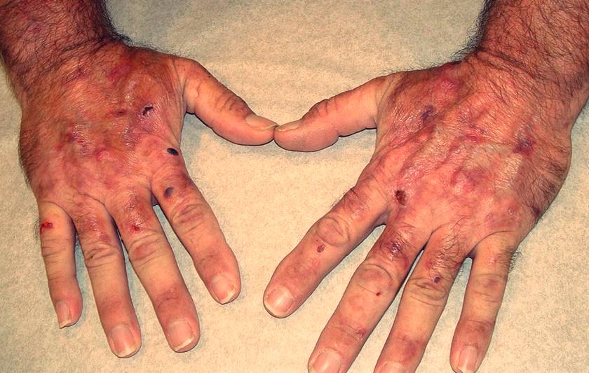
This accumulation causes pain in the skin with exposure to the sun (“freezing fire in the skin”), itching, redness, blisters, edema and even loss of substance in severe forms. These symptoms can also appear with artificial lights.

What are porphyrias and why do they occur?

The porphyrias are a group of at least nine genetic metabolic disorders described in the heme synthesis pathway. Heme is half of hemoglobin, along with globin. This molecule is responsible for transporting oxygen in the blood within the red blood cell or erythrocyte.

The heme group is synthesized partly in the liver and partly in the erythrocyte, which is why porphyrias are classified, depending on the mechanism that is altered, into hepatic and erythropoietic (erythrocytes or red blood cells).

In addition there are porphyrias not genetic, acquired due to liver failure or toxicity (1). A well-known one is the taking of oral tetracyclines (antibiotics) in some very susceptible patients.

Classification of porphyrias according to the organ in which abnormal porphyrins are synthesized

  • Erythrepoietic
    • Congenital erythropoietic porphyria (CEP)
    • Erythropoietic protoporphyria (EPP)
  • Hepatic
    • Preliminary cutaneous porphyria (PCT)
    • Hematoerythropoietic porphyria (HEP)
    • Porphyria variegata (PV)
    • Hereditary coproporphyria (CPH)
    • Acute intermittent porphyria (AIP)
    • Porphyria due to ALA dehydratase deficiency (Pd-ALAD)

How common are porphyrias?

Porphyrias are considered rare diseases. In the United States, they impact fewer than 200,000 people.

In Europe, the prevalence of the three most common forms of porphyria, cutaneous Tarta, acute intermittent and erythropoietic, is 1 in 10,000, 1 in 20,000 and 1 in 75,000, respectively.

Congenital erythropoietic porphyria, also called Günther's disease, is extremely rare, with an incidence of approximately 1 per million inhabitants.

the Nework European porphyria , a highly recommended page if you want to delve deeper into the topic and look for porphyria specialists in Europe, estimates about 335 patients diagnosed in 3 years in 11 countries (1,2).

How does a porphyria manifest?

Photosensitivity occurs in all porphyrias except acute intermittent porphyria and ALA dehydratase deficiency porphyria. This photosensitivity manifests itself in two ways (1):

  • Acute photosensitivity syndrome: This is pain, burning sensation and itching with sun exposure. Then redness and edema of the skin appear, like a disproportionate sunburn. This image is due to the accumulation of PpIX in the cells.
  • Skin fragility syndrome: It is not so acute, it is latent, and it is the appearance of erosions, blisters and cysts on the skin with minimal trauma or sun exposure.

Is there a common mechanism in porphyrias and photodynamic therapy?

Yes it exists. In a study made public in 2016 on neuron cultures, they discovered that the common mechanism is through ion channels (TRPA1 and TRPV1) that produce cellular damage through the activation of PpIX with ultraviolet and blue light (3).

They thus suggest the therapeutic opportunity of using medications that block these channels to prevent pain from sun exposure and skin damage

How is porphyria diagnosed?

The diagnosis is made under suspicion of symptom of the patient and determination of porphyrins in blood, urine or feces.

PpIX fluoresces when it accumulates in cells, and if we illuminate with a wood light or black light, widely used for diagnosis in dermatology, patients' teeth are fluorescent red (erythrodontics).

This is exactly the method we use to confirm that PpIX has accumulated in the cells we want when we do Photodynamic Therapy and we call it: “Fluorescence Diagnostics” (4). See next image:

Porphyrias: fluorescence diagnosis when illuminating with Wood's light

This image shows a basal cell epithelioma and how after inducing protoporphyrin IX inside, when illuminated with Wood's light, red-pink fluorescence can be seen due to its accumulation.

How can PpIX activation and porphyrin skin damage be avoided?

To understand it better I explain it in the following image:

Porphyrias: spectrum of electromagnetic radiation

The top part shows the spectrum of electromagnetic radiation, where we can see as the wavelength increases:

A) Ultraviolet light

Which damages the skin and produces cancerous and precancerous lesions. It's divided in:

1. Ultraviolet A (UVA): the one that makes us brown. 400 to 315 nanometers (nm)

two. Ultraviolet B (UVB): the one that burns us and more related to skin cancer. From 315 to 280 nm.

3. Ultraviolet C (UVC): 280 to 100 nm. It is not good either, but due to its wavelength it reaches the ground little and penetrates the skin little.

B) Visible spectrum

What we see, the colors. It's a variety.

1. blue: 380 to 427 nm.

two. Green: from 497 to 570 nm.

3. Yellow: 570 to 580nm.

Four. Red: From 600 to 780 nm.

C) Infrared

1. Above 800 nm, it no longer acts as a source of photostimulant.

Below is the absorption spectrum of protoporphyrin IX (PpIX), which is divided into:

A) Soret Band

The most toxic, because it is the highest on the graph. In the ultraviolet range.

B) Four Q bands

Less toxic, ranging from blue to red.

When does light cause the most damage in people with porphyrias?

The damage that porphyrin causes to the skin when exposed to the sun occurs mainly when the PpIX matches (5,6):

  • The ultraviolet range and the blue range: is the most toxic (around 400-420 nm)
  • Red light: 600-630nm: Why else is he in a less toxic band? Because the longer the wavelength, the greater the penetration into the skin. Red light penetrates 1 to 2 mm into the skin and thus does more damage than green or yellow light when there is a buildup of ppIX.

Conclusions: importance of protection against sunlight and other sources

The last line shows how Sunscreens blocking the electromagnetic spectrum. With this we now reach the two main conclusions of how NOT to activate PpIX:

TO) You should use a physical-chemical sunscreen (most on the market) or pure physical, protects from the entire spectrum. It is essential to verify that we are not using a chemical-only filter.

B) Other light sources can promote the PpIX, with lower intensity but reminiscent of LED lights (television, lamps), the light that passes through the window (mostly UVA) or other light exposures. They are mild, but added together they can activate PpIX.

I hope I have not been too technical and have reached the readers. I understand that knowledge of the mechanism is essential, since in general dermatologists insist on protection against ultraviolet rays, but in the case of porphyrias it is somewhat more complicated.

References

1 Ramanujam V. Diagnosis of porphyria-Part 1: A brief description of the porphyrias. Protocols Curr Hum Genet 2015; 86: 1-26.

2 www.porphyria.eu

3 Babes et al. Photosensitization in porphyrias and photodynamic therapy involves TRPA1 and TRPV1. J Neurscience 2016; 36: 5264-5278.

4 Fernández-Guarino M. Retrospective, descriptive, observational study of the treatment of various actinic keratoses with topical methyl aminolevulinate and red light: results in clinical practice and correlation with fluorescence imaging. Actas Dermosifiliogr 2008; 10: 779-787.

5 Heerfort IM. Noninvasively measured skin protoporphyrin IX predicts photosensitivity in patients with erythropoietic protoporphyria. BJ Dermatol 2016; 175: 1284-1289.

6 Teramura T. Prevention of photosensitivity with protection adapted to the spectrum of action for erythropoietic protoporphyria. J Dermatol 2018; 45: 145-149.

Gatos, alergias y enfermedades dermatológicas en humanos

gatos-enfermedades-piel-humanos-5752102-4788309-jpg-9166697

Conozca la vinculación que verdaderamente existe entre gatos, alergia y enfermedades dermatológicas en humanos a partir de la revisión de literatura científica de calidad.

Venerado por los egipcios, perseguido en la Edad Media, asociado a las brujas, la feminidad, la mujer, la libertad, la elegancia, la noche, los madrileños. Inspiradores de cuentos, fábulas, escritores, poetas y cineastas. El más exitoso con diferencia en las redes sociales. los gatos disponen algo.

¿Y para dermatología? ¿Qué sabemos los dermatólogos y la literatura científica sobre la vinculación entre los gatos y la piel? En esta publicación, desmitifico las ideas preconcebidas revisando literatura científica de calidad.

Dios creó al gato para ofrecer al hombre el placer de acariciar a un tigre

Victor Hugo

Gatos y humanos: una vinculación cada vez más intensa

El pequeño felino es hoy en día la mascota más popular En Europa, según datos de 2017, hay más de 100 millones de gatos en los hogares europeos, en comparación con 84 millones de perros.

Los gatos además ganan en Internet. Se cita en un Post de Nueva York Veces, que una imagen de un gato produce cuatro veces más tráfico en las redes sociales que la de un perro. Varios gatos comparten el podio de “influencers” con otros animales en las redes sociales.

Como parece que nos gustan los gatos, en este artículo resumo el enfermedades de la piel más frecuentess quien puede enfrentar el dueños de gatos con la finalidad de favorecer la convivencia con nuestro querido minino.

En este momento el gato es la mascota más popular

Vinculación entre gatos, alergia y enfermedades dermatológicas en humanos

1 tiña

Los gatos son un reservorio de la tiña, en realidad, son los animales con más tiña. Las tiñas son causadas por mushrooms dermatofitos que infectan la piel queratinizada (que no es mucosa), cabello y uñas.

Los dermatofitos que habitan en animales se denominan zoólicos, siendo el más común Mycosporum canis que habita en perros y gatos (1). El 90% de las tiñas en los gatos son causadas por este hongo (2).

the symptom de una tiña en un gato son semejantes a las del hombre, con la aparición de parches circulares en la piel con descamación y alopecia.

Los gatos además pueden ser portadores crónicos de Mycosporum canis, fundamentalmente los de pelo largo y los menores de un año (1,2). Estos gatos sin síntomas de tiña pueden ser transmisores.

The diagnóstico se realizan en base a los signos clínicos del animal y el cultivo de las escamas, a pesar de todo, no siempre es positivo pese a presentarse y no se detecta hasta en un 28% de los gatos infectados (3).

Lo mismo ocurre con la tiña en el ser humano, el hongo es exigente y en los cultivos no siempre se cultiva. Como opción al cultivo, se puede realizar un examen microscópico directo de las escamas para visualizar el hongo y evaluar la piel infectada con una luz de Wood (una luz negra) bajo la cual el dermatofito emite fluorescencia (3). Precisamente igual que en los humanos.

the treatment Se realiza con baños o geles antifúngicos, de eficacia limitada en gatos debido a la menor penetración en el pelo (2), por lo que cuando afecta al pelo es recomendable añadir además tratamiento oral al animal. Ambos tratamientos se mantienen durante unas 10 semanas.

En los humanos suelen requerir un tratamiento semejante, y se debe agregar un tratamiento oral siempre y cuando el cabello se vea afectado.

La infección es adquirida de forma general por contacto directo con el gato y más raramente por contacto con las escamas infectadas.

La mejor estrategia de prevención es evitar el contacto con el gato infectado, a pesar de todo, a veces el gato no muestra signos y el diagnóstico se realiza mediante una persona infectada. Después debemos llevar a nuestro gato a un veterinario (1).

Las esporas de Mycosporum canis son muy resistentes en el medio ambiente, por lo que descontaminación domiciliaria (dos).

Debe aspirarse y limpiarse mecánicamente hasta que no se vea ningún pelo. Es fundamentalmente importante desinfectar el lugar donde duerme, yace o vive el gato, así como los cojines o mantas que usa. Esta desinfección es fundamental en las trampillas para gatos.

Muchos desinfectantes están etiquetados como “antifúngicos” y son capaces de matar las esporas de los hongos dermatofitos, los más efectivos son los azoles, y no olvidemos la lejía doméstica, que diluida 1:10 en agua es un gran desinfectante (dos).

Deben aplicarse en todas las superficies, a la hora de lavar la ropa en la lavadora y además se pueden rociar. Existen en todas las variantes para estos usos en el mercado.

2 Alergia

Tema controvertido. Se sabe desde hace mucho tiempo que la exposición a los ácaros del polvo doméstico predispone al desarrollo posterior de alergia a otros alérgenos domésticos. De esta manera, los estudios encuentran que los niños “de interior” en las ciudades son más susceptibles a ser alérgicos.

Por el contrario, esta asociación no se ha encontrado en niños expuestos a animales de granja (4). Aún cuando la lógica parece indicar que el riesgo de sensibilización a alérgenos de perros o gatos aumentaría con la exposición a ellos, los estudios han demostrado todo lo contrario (4,5).

En un estudio consistente, Custovic et al (5), muestran que la prevalencia de sensibilización o La alergia a los gatos disminuye con la exposición. y que tener un gato se asocia con menos oportunidades de ser alérgico al animal.

Esta protección no se ha encontrado en dueños de perros adultos. La alergia a los gatos se evalúa midiendo los niveles de inmunoglobulinas específicas en la sangre contra el gato e introduciendo los antígenos pinchados en el brazo (“prueba de púa”).

Los pacientes con alergia a los gatos generalmente son polialérgicos y sensibles a otros alérgenos, como pólenes, perros o ácaros.

No se han encontrado diferencias en la gravedad de la alergia o el asma, tenga o no un gato (4), dicho de otra forma, tener un gato no aumenta la gravedad de los síntomas.

Parece que la exposición crónica a un gato si es alérgico a los gatos puede tener un “efecto protector” en niños y adultos al modular la solución inmunitaria (4).

Si deben ser fundamentalmente cuidadoso the pacientes alérgicos a los gatos que no disponen gato, puesto que la exposición ocasional a alérgenos de gatos puede provocar reacciones alérgicas y respiratorias con exacerbación del asma.

3 Dermatitis atópica

Los médicos a veces aconsejan a las familias con dermatitis atópica, ya sean niños o adultos, que se deshagan de sus mascotas peludas.

No hay evidencia científica que respalde esta recomendación. En los análisis científicos de máxima calidad en los que se revisan todos los trabajos publicados, no se demuestra que tener un animal en casa, inclusive desde el nacimiento, cause algún daño, ni siquiera si tiene dermatitis atópica.

Al mismo tiempo, las mascotas parecen tener efecto protector. Peluchi et al (8), en un estudio ni más ni menos que en 71.721 pacientes, encontraron que tener un perro reduce el riesgo de tener dermatitis atópica en un 30% y tener un gato no lo aumenta.

Entonces médicos y usuarios, los trabajos de mejor calidad en las mejores revistas son claros, como dicen los propios autores: “No hay razón para patear al gato”.

Gatos y enfermedades de la piel en humanos: conclusiones

Finalmente podemos decir que tener un gato no empeora the atopic dermatitis, ni el allergyInclusive podría tener un efecto protector que además podemos extender a tus amigos perros. Al contrario, son buenos transmisores de tiña.

References

1 Monod M, Fratti M, Mignon B, Baudraz-Rosselet. Dermatofitos transmitidos por animales domésticos. Rev Med Suisse 2014; 10: 749-5.

2 Frymus T, Gruffydd-Jones T, Pennisi MG, et al. Dermatofitosis en gatos: directrices ABCD sobre prevención y tratamiento. Revista de Feline Med Surg 2013; 15: 598.

3 Cafarchia C, Romito D, Sasanelli M, et al. La epidemiología de las dermatofitosis canina y felina en su otra Italia. mycoses 2004; 47: 508-513.

4 Liccardi G, Martín S, Lombardero M, et al. Respuestas cutáneas y serológicas a alérgenos de gatos en adultos expuestos o no a gatos. Neumología 2005; 99: 535-534.

5 Custovic A, Simpson BM, Simpson A, et al. Reducción de la prevalencia de sensibilización en gatos con alta exposición a alérgenos de gatos. J Allergy Clin Immunol 2001; 108: 537-9.

6 Williams HC, DJ de Grindlay. ¿Qué hay de nuevo en el eccema atópico? Un análisis de revisiones sistemáticas publicadas en 2007 y 2008. Parte 2. Prevención y tratamiento de enfermedades. Clin Exp Dermatol 2009; 35: 223-227.

7 Langan SM, Flohr C, Williams HC. El papel de las mascotas peludas en el eccema: una revisión sistemática. Arch Dermatol 2007; 143: 1570-7.

8 Peluchi C, Galeone C, Bach JF y col. Exposición de mascotas y riesgo de dermatitis atópica en la edad pediátrica: un metanálisis de estudios de cohortes de nacimiento. J Allergy Clin Immunol 2013; 132: 616-22.

10 benefits of melatonin on the skin

beneficios-melatonina-piel-2621932-7098942-jpg-6976931

Learn 10 of the main benefits of melatonin on the skin and its use in dermatology.

Melatonin is a hormone produced by the pineal gland whose secretion is influenced by the sleep-wake or light-dark cycle. Its main function in the body is to regulate sleep cycles.

The production of melatonin by the human body decreases with age. It is known that its supplementation has anti-aging effect, improving the quality of the skin and protecting it from ultraviolet radiation and other environmental damage such as tobacco.

What is melatonin?

Melatonin is a hormone produced by the pineal gland whose secretion follows a circadian rhythm, influenced by the cycles of light and darkness. It has a wide variety of properties at the cellular level including a high antioxidant power.

Like the entire body, the skin undergoes the oxidation and aging process, caused by internal and external factors, such as ultraviolet light or the production of free radicals. Melatonin levels increase at night and in mammals it has been shown to modulate immune response, body weight, anti-jet-lag effects, reduce the toxic effects of chemotherapy and be an anti-cancer agent.

The benefits of melatonin on the skin are varied. It has anti-aging effects and is a powerful antioxidant, useful as adjuvant in various dermatoses.

Melatonin in dermatology can be used as an oral supplement at bedtime or safely formulated into creams for topical use.

10 benefits of melatonin on the skin and main uses in dermatology

  1. Anti-aging and anti-wrinkle.: Melatonin is a sunscreen and has an anti-wrinkle effect. Its main action focuses on the keratinocytes and fibroblasts of the skin, protecting them from damage from ultraviolet radiation. It has also been shown to reduce the production of free radicals in the skin (1,2).
  2. Sun tanning: its intake before sun exposure reduces the risk of sunburn and its application in a 0.5% cream is the most powerful concentrate capable of removing the erythema or redness caused by a sunburn (ultraviolet light) (3).
  3. Atopic dermatitis: Preliminary studies have shown the beneficial effects of melatonin in patients with atopic dermatitis, both in cream and supplemented form (4). Despite everything, it should not be routinely recommended in cases where the patient also has asthma, since it can increase bronchial reactivity (5).
  4. Skin damage from radiotherapy (Radiodermatitis): Melatonin has been shown to have a radioprotective effect, reducing skin damage caused by radiotherapy and increasing its therapeutic effect. Treatment must be prolonged (6).
  5. Spots on the skin (melasma, solar lentigines): melatonin is a promising and safe treatment to reduce skin imperfections. Its mechanism of action is based on the inhibition of the action of tyrosine oxidase on the skin, responsible for the appearance of spots or pigmentation (7).
  6. Androgenic alopecia: It can be used topically or orally as a complementary treatment for androgenic alopecia and other forms of alopecia due to its powerful antioxidant action (8).
  7. Squamous cell carcinomaRecent in vitro studies have found that melatonin induces squamous cell carcinoma cell death (9). Its use in this indication is not widespread, but it is in the context of being a powerful sunscreen that predisposes to the development of skin cancer, this being one of the main benefits of melatonin on the skin.
  8. smoker's skin (skin and tobacco) and postmenopausal women: supplemented melatonin has anti-aging action in this group of patients, reducing wrinkles and imperfections and increasing skin elasticity. It is capable of reversing the changes of tobacco on the skin (10).
  9. Vitiligo: in vitiligo we find a sensitivity of melanocytes to oxidative stress that leads to their destruction. Melatonin has a powerful antioxidant action on the skin and a protective role for the melanocyte, which is why it can be associated with other treatments (11).
  10. Melanoma: Melatonin has action against cancer, so it can be beneficial as an adjuvant treatment in breast, liver, prostate or melanoma cancer.

Bibliography

1. Kleszczynski K, Fisher T. Melatonin and human skin aging. Dermatoendocrinol 2012; 4: 245-252.

2. D-Day, Burgees CM, Kircik LH. Evaluation of the potential role of topical melatonin in an anti-aging skin regimen. J Drugs Dermatol 2018; 17: 966-969.

3. Banha E, Elsner P, Kistler GS. Suppression of UV-induced erythema through topical treatment with melatonin (N-acetyl-5-methoxytryptamine). A dose response study. Arch Dermatol Res 1996; 288: 522–6.

4. Shi K, Lio PA. Alternative treatments for atopic dermatitis. Am J Dermatol 2018; E-publishing ahead of print.

5. Marseglia L, D'Angelo G, Manti T, et al. Melatonin and atopy: role in atopic dermatitis and asthma. Int J Mol Sci 2014: 15: 13482-13493.

6. Shabeed D, Najafi M, Musa AE, et al. Biochemical and histopathological evaluation of the radioprotective effects of melatonin against skin damage induced by gamma rays. Curr Radiopharm 2018; E-publishing ahead of print.

7. Juhasz MLV, Levin MK. The role of systemic treatments for skin lightening. J Cosmet Dermatol 2018; 17: 1144-1157.

8. Hatem S, Nasr M, Moftah NH, et al. Vitamin C-based melatonin nanovehicles for the treatment of androgenic alopecia. Eur J Pharm Sci 1028; 122: 246-253.

9. Kocyigit A, Guler EM, Karatas E, et al. Dose-dependent proliferative and cytotoxic effects of melatonin on human squamous cell carcinoma and normal skin fibroblast cells. Mutat Res 2018; epub before printing.

10. Sagan D, Stepniak J, Gesing A, et al. Melatonin reverses increased oxidative damage to membrane lipids and improves the biophysical characteristics of the skin in former smokers. Ann Agric Enviton Med 2017; 24: 659-666.

11. Patel S, Rauf A, Khan H et al. A holistic review on the autoimmune disease vitiligo with emphasis on causal factors. Biomed Pharmacother 2017; E-publishing ahead of print.

12. Srinivasan V, Pandi-Perumal SR, Brzezinski A, et al. Recent immune drug discovery by Pat Endocr Metab 2011; 5: 109-123.

Beneficios del yoga en la piel y rejuvenecimiento

beneficios-del-yoga-en-la-piel-6974172-4705463-jpg-4493782

El yoga es una práctica milenaria con beneficios para el cuerpo y la mente. La práctica del yoga ha demostrado beneficios para la salud en trabajos científicos de calidad. continua leyendo esta publicación para conocer específicamente los beneficios del yoga en la piel.

La práctica de la paciencia, perseverancia, autorrealización y ahimsa (no violencia hacia uno mismo) mediante asanas (posturas), pranayamas (respiración) y meditación, parece que podemos rejuvenecer.

La palabra yoga tiene dos significados, unión, con uno mismo y con el cosmos. Se puede establecer como las técnicas de mejora del ser humano a nivel físico, mental y espiritual.

Origen del yoga y beneficios para la salud

La antigua cultura del yoga debe su evolución a los antiguos sabios que desarrollaron sus enseñanzas y las transmitieron a sus discípulos a través del contacto directo con el maestro.

Su origen se remonta a la antigua civilización del Indo en el tercer y segundo milenio antes de Cristo. Sus enseñanzas fueron transcritas a los textos clásicos de los “Vedas” en los que el yoga se menciona por primera vez y adquiere una base establecida.

El yoga se ha extendido y es una práctica universal dentro de la cual hay diferentes caminos, inalcanzables en este artículo, pero de manera muy breve podemos hablar de Hatha yoga (yoga físico y sus variantes) y Raja yoga (yoga mental) (1).

Para algunos autores, el yoga es un estado de ánimo, una forma de calmar la mente y alcanzar la paz interior, que se cumple mediante posturas, “asanas” y técnicas de respiración, “pranayama” (2).

At literatura científica de calidad encontramos más de 4000 trabajos dedicados al estudio del yoga y su efecto a nivel psicológico y físico. Podemos ver que la mayoría de ellos se centran en trastornos neurológicos, artritis, manejo del dolor, emociones o cualidades cognitivas. Pero en nuestro caso nos vamos a centrar específicamente en los estudios que abordan los beneficios del yoga en la piel.

Beneficios del yoga en la piel

Anti arrugas

La vida yogui es antiarrugas: y hay evidencia molecular que lo prueba. La piel expresa nuestra apariencia y nuestra edad y tiene sentido que lo haga en paralelo a los órganos internos (3). La práctica regular de asanas y pranayamas reduce la actividad de la enzima superóxido dismutasa (SOD), una de las principales implicadas en el envejecimiento, y lo hace regulando la expresión de genes (4).

Al mismo tiempo, aumenta la expresión de moléculas reparadoras reconocidas (COX-2 y bcl-2) y antiinflamatorias (IL6, TNF), y lo hace mejor que el ejercicio extenuante (5).

Otras vías involucradas en el envejecimiento además disminuyen su actividad (AGE, productos finales de glicación avanzada), aumentan el flujo sanguíneo y mejoran el metabolismo general de la piel y los tejidos (3).

Probablemente la práctica de una actitud yogui con su mejora en la calidad de vida ayude a estar menos oxidado y a ser más joven.

Expresión facial

El yoga regula la expresión facial: los practicantes de yoga habituales expresan menos emociones negativas en sus rostros en situaciones negativas. El yoga reduce la “excitación”, que es la respuesta del cuerpo a las emociones negativas o al estrés y consiste en la aparición de sudor, taquicardia, respiración acelerada.

Los pacientes estudiados, al mismo tiempo de tener menos expresiones corporales de excitación a las emociones negativas, además reportaron menos experiencia emocional negativa a los estímulos negativos (6).

Acné excoriado y tricotilomanía

Otro beneficio del yoga en la piel se puede hallar en el tratamiento del acné excoriado y la tricotilomanía.

El acné excoriado es la manipulación de las lesiones del acné, común en mujeres adolescentes, que produce cicatrices e inclusive infecciones locales.

La tricotilomanía es otro hábito que se encuentra en los niños pequeños, que arrancan mechones de cabello en momentos de estrés.

Ambos se deben a conductas repetitivas centradas en el cuerpo y bajo las cuales encontramos procesos de ansiedad o adaptación. Se ha demostrado que la práctica del yoga agrega beneficios a los tratamientos farmacológicos y psicológicos para estas afecciones (7).

El yoga facial es antiarrugas

En un estudio reciente difundido en una revista de dermatología de calidad, un grupo de pacientes que realizó yoga facial durante 20 semanas encontró una reducción moderada de las arrugas en el tercio medio y el tercio inferior. El programa consiste en practicar los ejercicios durante 30 minutos todos los días de la semana a 2 o 3 veces por semana. Su método de acción puede deberse al entrenamiento de los músculos faciales (3).

Conclusiones sobre los beneficios del yoga en la piel.

En mi opinión, el yoga no sustituye a otros métodos como una buena crema antiarrugas con ácido retinoico u otras técnicas (toxina botulínica, rellenos, peelings, etc.), y todavía faltan más estudios de calidad, aún cuando puede ayudar (fundamentalmente si produce bienestar físico y mental).

Bibliography

1. Danilo Fernández. Claves del yoga: teoría y práctica. XIX Edición 2017. La Liebre de Marzo, SL.

2. Cristina Brown. La biblia del yoga. 1ª Edición 2015. Gaia Ediciones SL

3. Beri K. Respirar para una piel más joven: “revertir el mecanismo molecular del envejecimiento de la piel con el yoga”. Future Sci OA 2016; 2: 122.

4. Saatcioglu F. Regulación de la expresión génica por el yoga, la meditación y prácticas asociadas: una revisión de estudios recientes. Asian J Psychiatr 2013; 6: 74-77.

5. Vijayaraghava A, Doreswamy V, Narasipur OS, et al. Efecto de la práctica del yoga sobre los niveles de marcadores inflamatorios luego de ejercicio moderado y estenoso. J Clin Diagn Res 2015; 9: 08-12.

6. Mocanu E, Mohr C, Pouyan N y col. Razones, años y frecuencia de la práctica del yoga: efecto sobre la reactividad de la respuesta a la emción. Fornt Hum Neurosci 2018; 12: 264.

7. Torales J, Barrios I, Villalba J. Terapias formas de el trastorno de excoriación (pellizcarse la piel): una breve actualización. Adv Mind Body Med 2017; 31: 10-13.

8. Alam M, Walter AJ, Geisler A, et al. Asociación del ejercicio facial con la aparición de envejecimiento. JAMA Dermatology 2018; 154: 365-367.

Skin care during chemotherapy

cuidados-piel-quimioterapia-1-5849537-5944856-jpg-7830693

Learn some tips on skin care during chemotherapy focused on addressing the most common skin side effects.

Cancer treatment is a medical and research challenge. The chemotherapy Traditionally, it works by inhibiting phases of the cell cycle of cancer cells, but it can also affect healthy cells. The skin is one of the organs most affected, with the possible appearance of dermatitis, rashes, canker sores, spots, nail alterations and other injuries.

Conventional or traditional chemotherapy continues to be used to control tumors. Advances in cancer treatment and patient survival make it necessary for clinicians to know how to manage chemotherapy reactions on the skin and advise patients. In the following text I present some guidelines for managing side effects and skin care during chemotherapy.

Skin Side Effects of Chemotherapy

The classification of cutaneous side effects of chemotherapy is standardized by the National Cancer Institute's Common Terminology Critherapy for Adverse Events (CTCAE) (1) in a common tool for all side effects of chemotherapy and that allows unifying the evaluation of performed by oncologists, dermatologists, nurses and other clinicians (available clicking here and summarized in Table I). These effects they do not always occur, this is a classification of all possible skin conditions when a patient is treated with chemotherapy (2).

Side effects of chemotherapy on the skin.

The majority of chemotherapy reactions on the skin are managed in oncology services, who are aware of its effects and with nursing professionals specialized in them. Only occasionally they notify the dermatologist, when the skin lesions cause diagnostic doubts or when they are more severe or do not respond to the usual treatments. In the following text I will explain the most frequent effects and general advice for their management (3). Alopecia due to chemotherapy would be its own content due to its entity, so in this text I will only address the effects on the skin.

Dermatologists study these effects and skin care during chemotherapy, since the priority in these patients is the response of the tumor to the treatments received, which means that sometimes, we are “companions” during the process.

Side effects and skin care during chemotherapy

1. Dry skin or cutaneous xerosis

It is one of the most common side effects of chemotherapy on the skin. The creams that I usually recommend to alleviate it are emollients and fats, with soothing and anti-inflammatory substances (calamine, vitamin PP, oats, omegas, etc.). The prototype of creams would be those used for children with atopic dermatitis, although there are specific lines on the market studied for oncology patients.

Some moisturizing creams contain keratolytic agents, which are added to also treat peeling. Above all, urea is used in concentrations from 3 to 30%, and lactic acid, salicylic acid and even glycolic acid can also be used. I recommend trying them first in low concentrations, as they are especially useful on dry skin with slight peeling, but at high concentrations they can cause irritation and itching on sensitive skin.

It is also important to use anti-aging facial care with caution, since very common substances in these products can irritate, such as retinoic acid or retinol, glycolic acid and other alpha and beta-hydroxy acids. Perhaps the most appropriate thing is to use “anti-aging” substances such as vitamin C, vitamin B3 or B5, melatonin, hyaluronic acid, resveratrol or coenzyme Q10 for the face, which have a low potential to irritate.

Regarding washing the skin, you can opt for shower oils or “soapless soaps” or “syndet” that moisturize the skin during the shower.

2. Pruritus or itchy skin

Itchy skin during chemotherapy can be due to several causes, including the drugs it contains. I emphasize that in this case we are talking about itching without lesions on the skin, when itching appears on the skin with lesions the treatment is that of the lesions. The approach, in my opinion, is to achieve skin that is as comfortable and hydrated as possible, with the measures in the previous point.

In addition, you can use hydrating or soothing baths, effective at night, when the skin tends to itch the most, to facilitate a night's rest, and oral antihistamines. Antihistamines are simple treatments that serve to relieve itching, and if they are sedative, facilitate sleep. There are more powerful oral treatments (in pills) and also anti-itch creams and you can even apply corticosteroid creams to the most itchy areas.

3. Dermatitis and eczema

Skin care during chemotherapy | Eczema.

Intense dryness of the skin and other added factors, such as chemotherapy or some irritant, can produce true inflammation of the skin, in the form of dermatitis and eczema. Management is with emollients or moisturizing creams that do not sting; it may be necessary to use pure Vaseline for a few days to avoid stinging. Corticosteroids in cream are added twice a day, which are effective in controlling dermatitis. In severe or extensive cases, oral corticosteroids and oral antihistamines can be added to control itching.

The measures on skin care during chemotherapy in points 1 and 2 are applicable in patients with dermatitis as well.

4. Thrush or mucositis

The appearance of thrush or mucositis during chemotherapy cycles has a significant impact, since it causes a lot of discomfort and makes eating difficult, in these patients where maintaining general condition is important (5). The term mucositis refers to inflammation of the mucous membranes, the most frequently affected is the oral mucosa, and it can also occur in the genitals.

Mucositis subsides in a few days and in the meantime, it can be treated with mouthwashes with anti-inflammatories (tetracyclines, corticosteroids) and anesthetics. Its management consists of relieving the patient. Anesthetics in the mouth can also be applied before eating, to avoid pain and facilitate feeding. There are also mouthwashes that moisturize the mucosa, and hyaluronic acid gels and other components to apply directly to canker sores. They act by protecting the skin and promoting healing. The patient must be monitored and if it is more intense or does not resolve with treatment, it could be a fungal superinfection or, more rarely, a herpes virus.

There is clearly no way to prevent this adverse effect, keeping the mucosa hydrated and vitamin supplements, especially A, important for the mucosa, could be used.

5. Alterations in the nails

Nail fungus. Possible side effect of chemotherapy.

Fragmentation, lines and striations (onycholysis) may appear in the nails during chemotherapy. It has been described with chemotherapy drugs such as taxanes, cyclophosphamide, doxorubicin, capecitabine, etoposide and 5-fluorouracil (2,3,5). For its management, it is advisable to avoid irritants to the nails, such as frequent contact with water and trauma. To carry out household chores, you can use plastic gloves with cotton gloves underneath (they are special and are found in pharmacies).

Manicures should be avoided and nails should be kept short. Moisturizing the nails and the periungual area benefits the nails, and you can use general moisturizing creams or specific nail care creams, also in the form of specific polishes (not coloring nail lacquer polishes). This side effect can be prevented by using cold gloves during the chemotherapy infusion, which produce vasoconstriction and reduce the arrival of the chemotherapy to the nail matrix.

Sometimes what occurs is a darkening of the nail (hyperpigmentation), as occurs with 5-Fluorouracil, taxanes, cyclophosphamide, doxorubicin and capecitabine (1,2). This pigmentation can appear in bands or diffusely on the nail plate. This is a side effect that does not require treatment and does not cause discomfort. It persists months after finishing chemotherapy and disappears with the progressive growth of the nail.

Rarely, the alterations produced by chemotherapy in the nails predispose to infection of the nail itself or the surrounding area (perionyxis). In this case, management by a dermatologist may be required and sometimes a culture may be performed to identify the infectious agent and be able to treat it.

6. Hyperpigmentation or appearance of spots on the skin

The appearance of spots on the skin and mucous membranes secondary to chemotherapy has a very varied clinical expression. It may be diffuse, with a tanned appearance, mottled, flagellated (lashed), serpentine, only in areas of flexion, or on the palms and soles (2,3).

It does not require treatment and subsides only months after finishing treatment. As general measures, within skin care during chemotherapy, sun protection is indicated in these patients and anti-blemish cosmetics can be used to treat and prevent their appearance. Hypothetically, pigment lasers could also be useful, and there is little about this use, remember that it is to treat an asymptomatic dermatosis that resolves on its own.

7. Photosensitivity

This is an exaggerated reaction to ultraviolet light from the sun and has been infrequently described with 5-Fluorouracil, Tegafur or Capecitabine (3). Erythema (redness) and intense inflammation occur in the area exposed to the sun. By default and to also avoid hyperpigmentation (spots), patients undergoing chemotherapy are protected from the sun and are encouraged to use sunscreen.

The recommended sunscreens are a factor 30 or higher, and use them before sun exposure. Let us remember that the sun protection index (SPF) measures only protection against ultraviolet radiation, which is responsible for phototoxic reactions. If we also want to protect ourselves from spots (hyperpigmentation), we must choose a filter that includes UVA protection, and if it also includes visible light, we protect ourselves from all light options that can darken the skin.

9. Palmar-plantar erythrodysesthesia

Skin care during chemotherapy: palmar-plantar erythrodysesthesia.

Also called hand-foot syndrome or acral erythema (4). It has been described with Capecitabine, doxorubicin, cytarabine, 5-fluorouracil and taxols (2,3,5). It consists of the appearance of a tingling sensation that can progress to burning in the palms and soles. Sensitivity to temperature and pain may be decreased and is accompanied by redness and swelling of the hands, and sometimes peeling. Erythrodysesthesia affects the palms more frequently than the soles and its severity varies.

To prevent the appearance of erythrodysesthesia, the way chemotherapy is administered is controlled by oncologists, and urea creams, non-steroidal anti-inflammatory drugs (NSAIDs) and hand cooling can be used during the chemotherapy infusion.

When it appears, treatment with powerful corticosteroids, local cold water compresses, and anti-inflammatories can be used. Other options are oral or topical calcium antagonists, anti-inflammatory COX-2 inhibitors, oral pyridoxine or topical heparin (2-4).

10. Maculopapular rash

This is a generalized rash from the treatment. It is common to any drug, and is treated according to its severity. Emollient creams, oral corticosteroid creams and oral antihistamines can be used for itching. In general, they are temporary and do not require discontinuation of treatment.

As part of skin care during chemotherapy, you can try to prevent these reactions by administering premedication before receiving the chemotherapy cycle, which includes antihistamines and oral corticosteroids (3).

To end and again, I would like to emphasize that this text is a transversal approach to all the possible effects of chemotherapy treatments and that They do not all occur at the same time with the same treatment. Prevention and treatment of chemotherapy side effects is essential in patients to avoid avoidable changes in administration protocols.

*On the other hand, if you are receiving radiotherapy, I recommend reading this article.

I hope it is useful to you.

References

1. https://ctep.cancer.gov/protocolDevelopment/electronic_applications/ctc.htm

2. Cury-Martins J, Eris APM, Abdalla CMZ, Silva GB, Moura VPT, Sanches JA. Management of dermatologic adverse events from cancer therapies: recommendations of an expert panel. An Bras Dermatol. 2020; 95:221-237.

3. Reyes-Habito CM, Roh EK. Cutaneous reactions to chemotherapeutic drugs and targeted therapies for cancer: part I. Conventional chemotherapeutic drugs. J Am Acad Dermatol. 2014;71: 203. e1-203.e12; perhaps 215-6.

4. Miller KK, Gorcey L, McLellan BN. Chemotherapy-induced hand-foot syndrome and nail changes: a review of clinical presentation, etiology, pathogenesis, and management. J Am Acad Dermatol. 2014; 71:787-94.

5. Balagula Y, Rosen ST, Lacouture ME. The emergence of supportive oncodermatology: the study of dermatologic adverse events to cancer therapies. J Am Acad Dermatol. 2011 Sep; 65:624-635.