
Removal of Pigmented Nevi with Laser: Effective and Safe Treatment
What are Pigmented Nevi?
Nevi, commonly known as moles, are growths on the skin formed by accumulations of pigmented cells called melanocytes. Although they are generally benign, it is important that any changes in their size, shape or color be evaluated by a dermatologist to rule out more serious problems such as melanoma.
Advantages of Laser Mole Treatment
The use of lasers for mole removal offers multiple advantages over traditional methods. This treatment has become a popular option due to its multiple benefits:
- Does not require anesthesia: Unlike other procedures that can be more invasive, laser treatment is virtually painless and does not require general anesthesia. In some cases, topical anesthesia may be used to minimize any discomfort.
- Non-surgical medical-aesthetic treatment: Being a non-invasive procedure, laser treatment does not involve cuts or sutures. This significantly reduces the risk of infections and other postoperative complications.
- Performed only by specialist doctors: Dermatologists and other highly trained medical professionals are the only ones authorized to perform this procedure, ensuring a high level of safety and effectiveness.
- Excellent cost-effectiveness ratio: Although it may seem like an expensive treatment, laser offers superior cost-effectiveness compared to other methods. The precision of the laser reduces the need for multiple sessions and additional treatments, saving time and money in the long run.
- Superior results: The results obtained with the laser are generally better in terms of aesthetics and functionality. The precision of the laser allows the mole to be removed with minimal scarring and without damaging the surrounding tissue.
Laser Treatment Process
Laser treatment for mole removal follows a well-defined process that guarantees its effectiveness and safety:
- Initial consultation: The first step is a consultation with the dermatologist, where the mole is evaluated and it is determined if laser treatment is appropriate. In this consultation, the risks, benefits and expectations of the treatment are discussed.
- Preparation: Before treatment, the area to be treated is cleaned and disinfected. In some cases, a numbing cream is applied to minimize any discomfort.
- Laser application: The dermatologist uses the laser to direct pulses of intense light toward the mole. The laser energy is absorbed by the pigmented cells, destroying them without damaging the surrounding tissue.
- Aftertreatment: After treatment, the treated area may be slightly red or swollen, but these effects are temporary. Aftercare is provided to speed healing and prevent infection.
- Tracing: Follow-up appointments may be scheduled to evaluate progress and determine if additional sessions are needed.
Considerations and Precautions
Although laser treatment is safe and effective, there are some considerations and precautions that should be taken into account:
- Type of skin: Skin color and type can influence the effectiveness of the treatment. Dermatologists adjust the laser settings to suit the individual characteristics of the patient's skin.
- Pre-existing medical conditions: Patients with certain medical conditions should inform their dermatologist before treatment to evaluate any possible contraindications.
- Aftercare: It is crucial to follow the aftercare instructions provided by the dermatologist to ensure proper healing and avoid complications.
Cost laser treatment for removal of moles
The cost of laser treatment for mole removal can vary depending on several factors:
- Number of sessions required: Some lesions can be eliminated in a single session, while others may require multiple sessions. This depends on the size, depth and location of the mole, as well as the patient's response to treatment.
- Initial evaluation by the dermatologist: It is essential that a dermatologist evaluate the lesion before proceeding with treatment. This evaluation allows us to determine the type of mole and the most appropriate treatment strategy.
- Technology used: The type of laser and the technology used will also influence the cost. More advanced and specific lasers to treat pigmented nevi may have a higher cost, but they also offer better results and greater safety.
To obtain a personalized quote for the removal of moles you must send photos via WhatsApp to the following numbers +54 911 5503 5126 and to +54 911 4079 3779
Treatment is performed by Dr. Ricardo Hoogstra in any of our 3 centers. You can schedule your shift via internet at any of our branches and attend directly to carry out a treatment.
Melanocytic nevi
The melanocytic nevi They are very common benign skin lesions that are found in almost the entire population. They are commonly known as moles and can vary in appearance and characteristics, but they share some common traits that make them identifiable.
Characteristics of Melanocytic Nevi
Melanocytic nevi can be flat or raised, asymptomatic, with well-defined edges, regular coloring, and frequently small in diameter (less than 6 mm). These moles can appear anywhere on the body and their color and shape can change over time.
Stages of development
Initially, the nevic cells are located between the epidermis and the dermis (dermo-epidermal junction) and are called junctional nevi or union. In this state, they are flat lesions of dark brown or black color. As time passes, groups of melanocytes (thecae) proliferate and extend into the dermis, becoming called compound melanocytic nevi.
Coloration of the Nevus
The color of a melanocytic nevus is related to the location of the pigment (melanin):
- Brown: If the majority of the pigmented cells are found in the epidermis.
- Dark brown or black: If they are located in both the epidermis and the superficial dermis.
- Bluish: If the melanocytes are located in the deep dermis, known as blue nevus.
Influence of Genetic and Environmental Factors
The number of melanocytic nevi that a person develops throughout their life is variable and is influenced by genetic and environmental factors, especially the degree of sun exposure to which the skin has been exposed.
Changes Throughout Life
Melanocytic nevi are dynamic proliferations that change throughout life. They can darken with sun exposure or during pregnancy and, in adulthood, they tend to progressively lose their pigmentation and can even disappear at advanced ages.
Types of Melanocytic Nevus
A) Congenital Melanocytic Nevus
The congenital melanocytic nevi They are present from birth and can vary significantly in size, from a few millimeters to covering a large part of the body surface. They are empirically classified into:
- Little ones: Less than 1.5cm.
- Intermediates: Between 1.5 cm and 20 cm.
- Giants: More than 20 cm.
These nevi present an increased risk of developing malignant melanoma, especially in cases of giant melanocytic nevi.
B) Acquired Melanocytic Nevi
The acquired melanocytic nevi They appear after birth and can vary in size, coloration and number. They are well defined and regular in color, and can be flat or raised. They are more common in areas exposed to the sun.
C) Atypical Nevi
The atypical nevi They are moles with irregular characteristics, including irregular borders, diffuse coloration, and larger size. These patients require periodic clinical and dermoscopic controls due to the increased risk of malignancy. Preventative surgical treatment is crucial, and many of these lesions must be removed and sent to a dermatopathologist for proper diagnosis.
Practical Tips for the Care of Melanocytic Nevi
Photoprotection
Sun exposure is one of the main factors that affect the appearance and changes in melanocytic nevi. It is crucial to use adequate sun protection:
- Sun creams: Apply sunscreen with a high protection factor (SPF 30 or higher) to all exposed areas, even on cloudy days.
- Protective clothes: Wear hats, sunglasses and clothing that covers the skin.
- Avoid hours of greatest radiation: Limit sun exposure between 10 am and 4 pm, when UV radiation is most intense.
Self-exploration
Performing regular skin self-examinations is essential to detect early changes in melanocytic naevi. Follow these steps:
- Check all the skin: Use a mirror to examine all areas of the body, including hard-to-see areas like the back and scalp.
- Check for changes: Pay attention to any changes in the size, shape, color or texture of existing moles and the appearance of new ones.
- ABCDE rule: Use this guide to identify worrisome features:
- A (Asymmetry): One half of the mole is not the same as the other.
- B (Edges): Irregular, uneven or poorly defined edges.
- C (Color): Variation of colors, including different shades of brown, black, pink or red.
- D (Diameter): Greater than 6 mm, although melanomas can be smaller.
- E (Evolution): Changes in size, shape, color or any other characteristic.
Regular Dermatological Controls
Scheduling regular visits to the dermatologist is crucial, especially if you have multiple nevi or atypical nevi. A dermatologist can perform a more detailed examination, using tools such as dermoscopy to evaluate lesions more precisely.
- Annual exams: At least once a year, or more frequently if you have additional risk factors.
- Track changes: The dermatologist can document and follow changes in the nevi to identify any worrisome changes early.
Treatment and Management
In some cases, treatment of melanocytic nevi may be necessary:
- surgical removal: Indicated in atypical nevi, giant congenital melanocytic nevi or in any nevus suspected of malignancy.
- Biopsy: It can be performed to obtain a definitive diagnosis if there is doubt about the nature of a nevus.
- active surveillance: For nevus that appears benign but in high-risk individuals, periodic check-ups can be scheduled.
Helios II
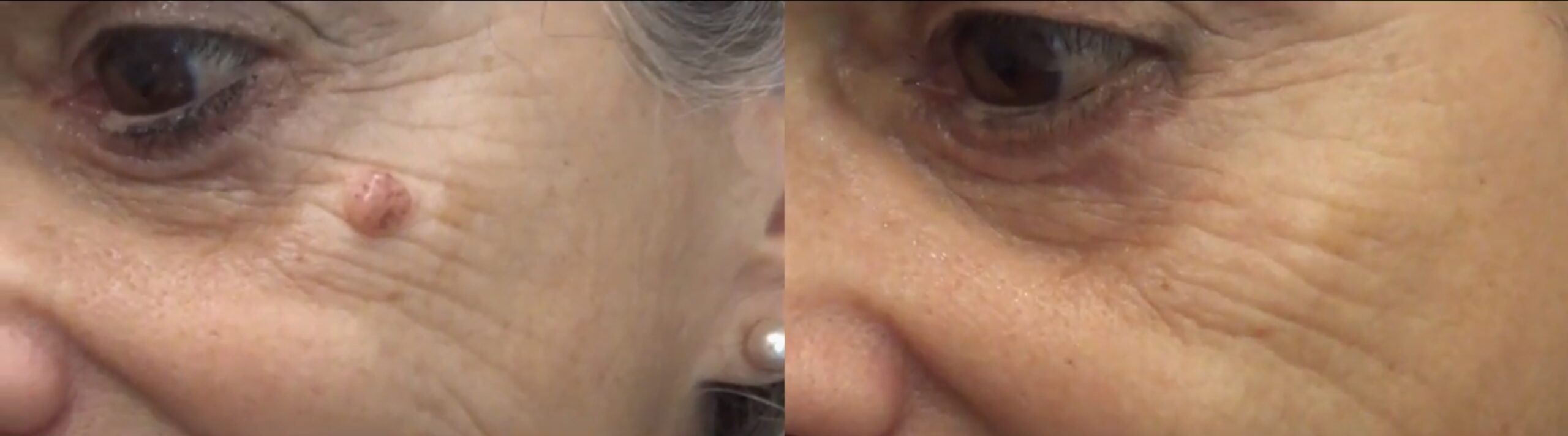
How is the treatment to remove moles?
Removing moles with a scalpel, whatever its size and number, leads to safe scars.
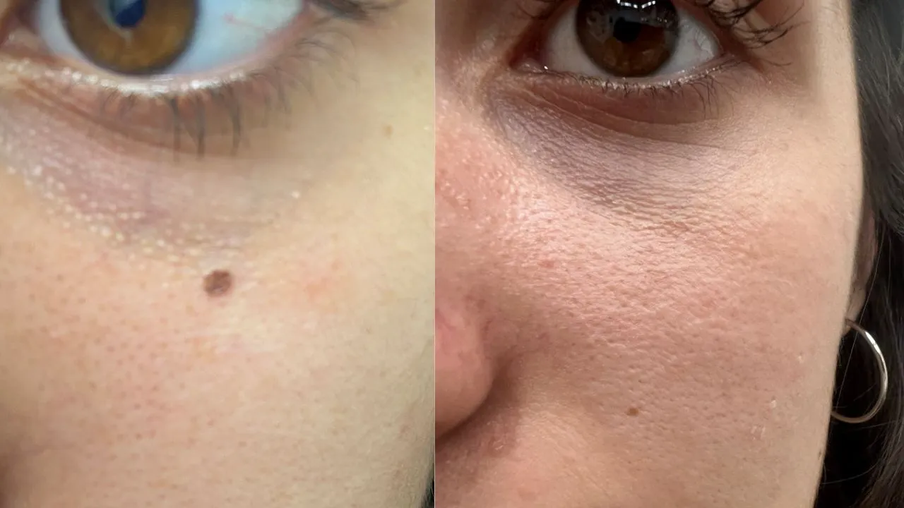
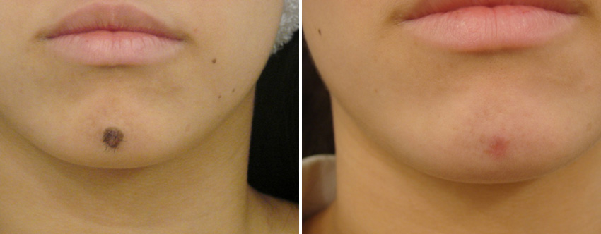
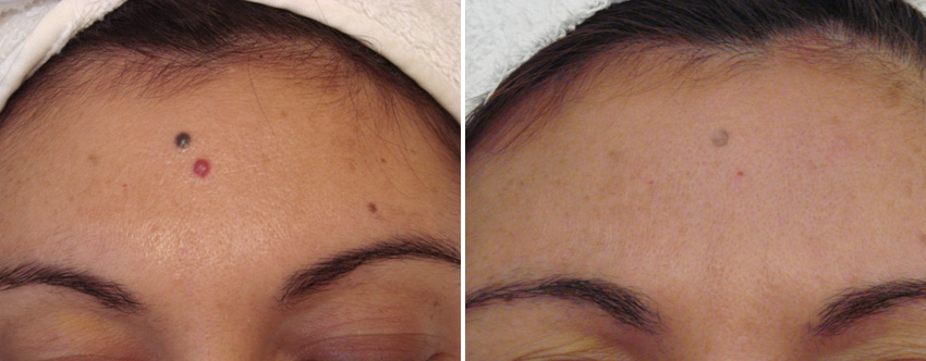
With the laser, a better aesthetic opportunity is obtained because the laser light is absorbed by the nevic pigment, consequently a heating and vaporization occurs that leads to the elimination of the nevus without damaging the neighboring tissue.
We could say that treatment with Helios II laser is more accurate. Patient satisfaction is high.
Recurrences are well accepted and easy to correct, therefore lasers generate more aesthetic results when it comes to removing moles for these purposes.
After more than 20 years using different laser technologies I have discovered that the Helios II Nd: YAG Q SWITCHED laser With dual pulse mode 1,064 and 532 nm it is the ideal instrument to remove moles and nevi like Ota's, quickly and safely, without scars and with very satisfactory aesthetic results.
Nevi are mostly benign skin lesions, however, prior to any laser treatment, a clinical, videoscopic and / or pathological assessment of the lesion must be performed. Most of the moles can be removed with Laser in a single session and the patient can immediately continue with his normal life, without taking any type of occlusive cure.
Advantages of treatment of moles with Laser Helios II:
• Does not require anesthesia.
• It is a non-surgical medical-aesthetic treatment performed only by specialist doctors.
• Normally with a single session the permanent elimination of the lesion is achieved.
• Excellent cost-effectiveness ratio.
• Results superior to those obtained with other procedures.
Most moles are completely benign and do not have any risk of malignancy, but all nevi must be surgically removed and sent for histopathological study that:
• It grows rapidly
• It has color and irregular edges
• Bleeds
• Frequently irritated
• They are in the friction zone as the sole of the foot
• They are very dark and are found in areas of difficult control, such as the scalp, perianal, etc.
• Large nevi
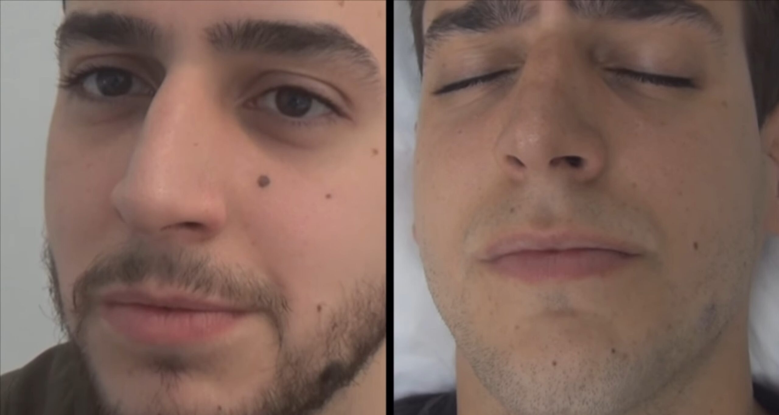
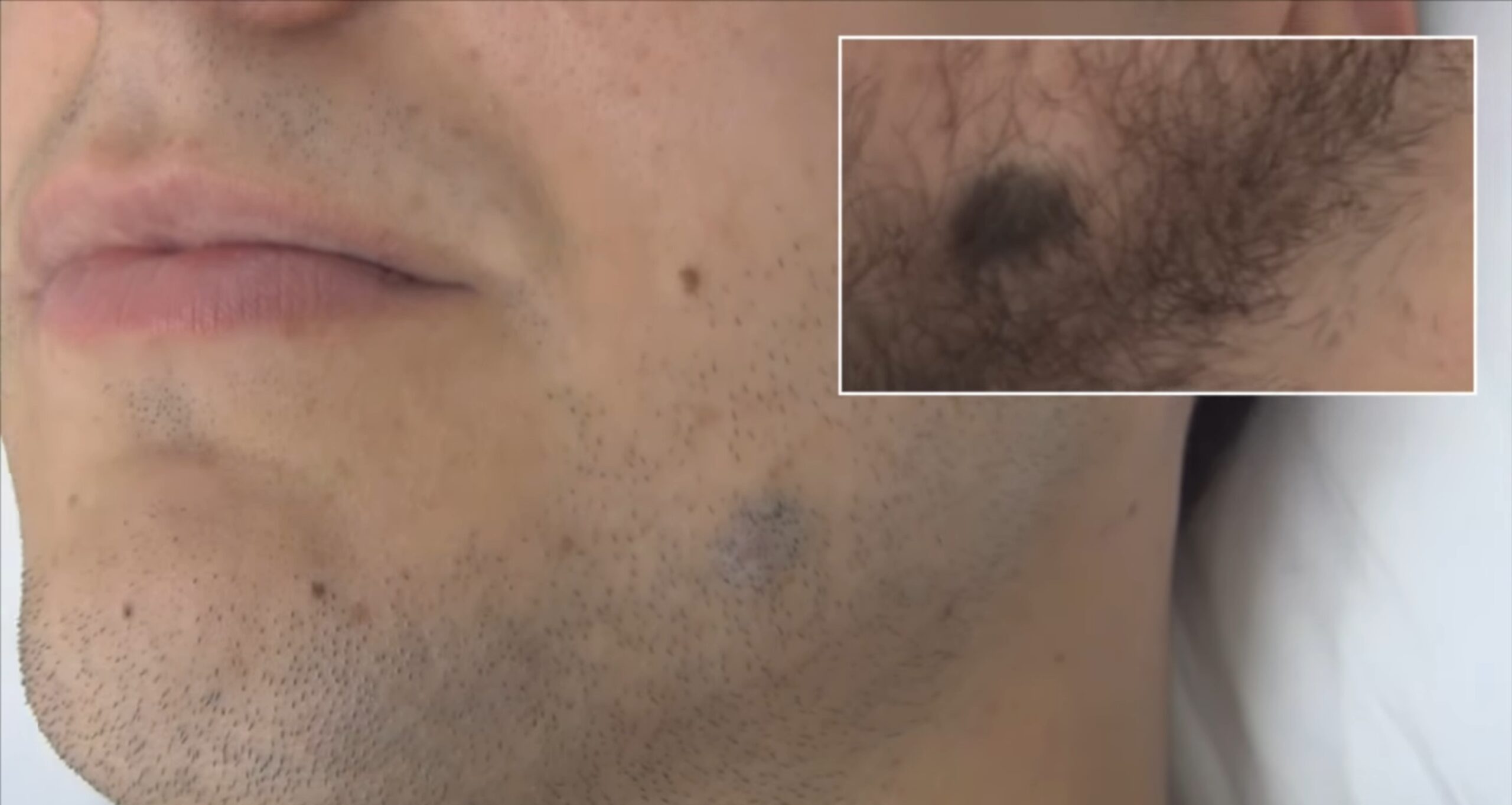
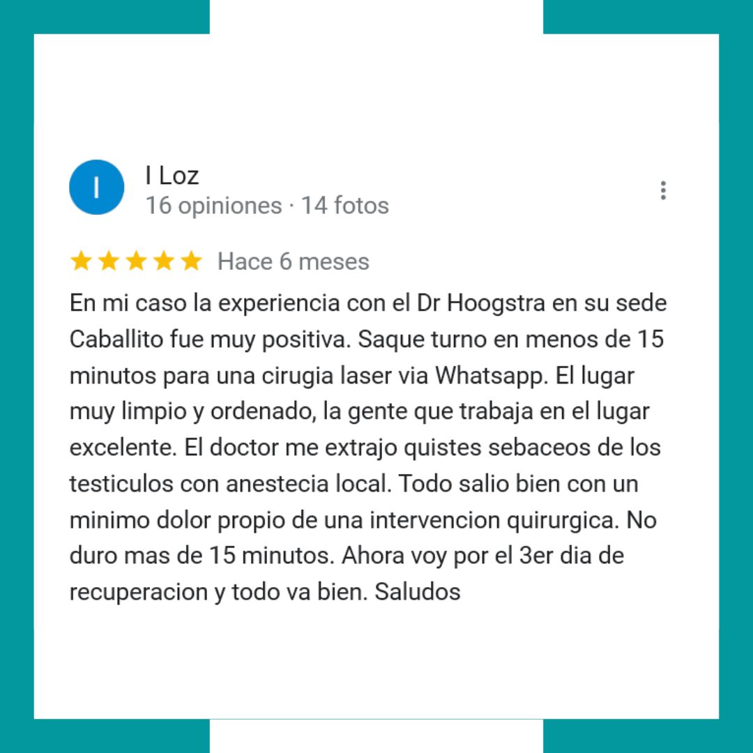
Patient testimonials
How do I make an appointment?
The most effective, safe and practical way to take a shift is via the internet.
If you have difficulty taking a shift online, payments and administrative issues.
You can call Monday through Friday from 8 a.m. to 8 p.m. (011) 4901-6690 (011) 4904-3434 (011) 4904-0880
You can take a turn and go directly to perform a treatment. If you have any questions, you can clarify it first with the same professional.
If you. If you have any medical questions, you can write to us in advance by sending an email to
Only medical consultations will be received here. This mail does not address inquiries about shifts, payments and administrative issues.
