
What are the perpendicular white lines?
On dermoscopy, the perpendicular white lines are short. discreet white lines oriented parallel and orthogonal (perpendicular) to each other and seen only under polarized light [1]. They are also known as polarizing white lines, short white lines, bright white lines, bright white stripes, chrysalis, pupae, and crystalline structures. Perpendicular white lines are a clue to a specific diagnosis that includes basal cell carcinoma (BCC) and some melanomas [2].
What do the perpendicular white lines look like through the dermatoscope?
Perpendicular white lines are only visible under polarized light. They appear as short, bright white lines and move as the dermoscopy lens is moved at different angles over the injury.
Perpendicular white lines in BCC
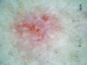
Superficial dermoscopy of basal cell carcinoma

Superficial dermoscopy of basal cell carcinoma
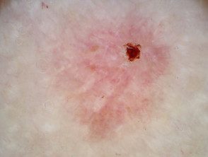
Superficial dermoscopy of basal cell carcinoma
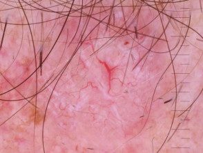
Basal cell nodular carcinoma dermoscopy
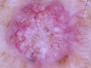
Basal cell nodular carcinoma dermoscopy
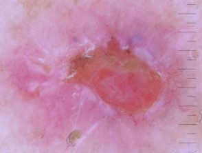
Basal cell nodular carcinoma dermoscopy
Perpendicular white lines in melanoma
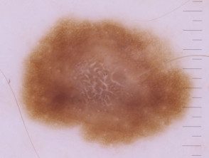
Dermoscopy of melanoma in situ
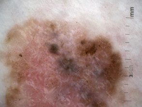
Dermoscopy of melanoma in situ
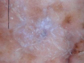
A nodular melanoma arising within a superficial spreading melanoma.

Amelanotic nodular melanoma
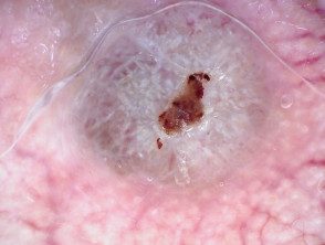
Nodular melanoma, Breslow 6.8 mm
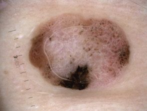
Nodular melanoma, Breslow 2.5 mm
polarized and non-polarized light
The following pairs of images demonstrate the differences seen in dermoscopy of perpendicular white lines under polarized and unpolarized light.
Polarized and unpolarized light in BCC
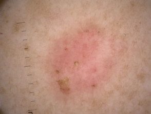
Non-polarized dermatoscopy view of BCC
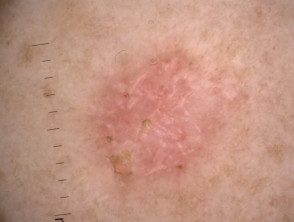
Polarized dermatoscopy view of BCC
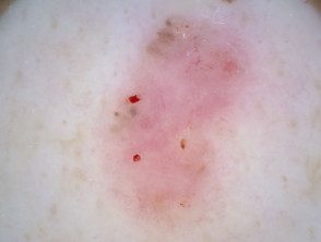
Non-polarized dermatoscopy view of BCC
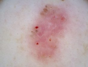
Polarized dermatoscopy view of BCC
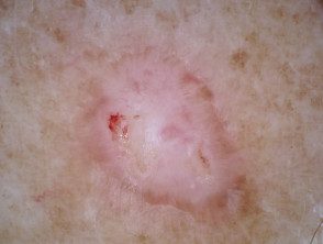
Non-polarized dermatoscopy view of BCC
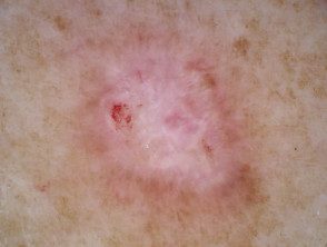
Polarized dermatoscopy view of BCC
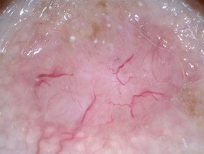
Non-polarized dermatoscopy view of BCC
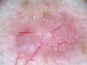
Polarized dermatoscopy view of BCC
Polarized and non-polarized light in melanoma
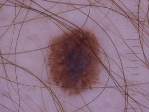
Perpendicular white lines are absent in melanoma in situ when viewed with non-polarized light dermoscopy.
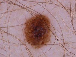
Perpendicular white lines are visible under polarized light dermoscopy of a melanoma in situ
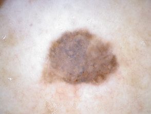
Melanoma in situ view, non-polarized dermatoscopy
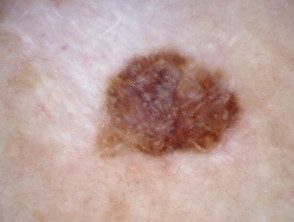
Melanoma in situ, polarized dermatoscopy
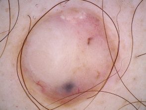
Non-polarized dermatoscopy view of nodular melanoma
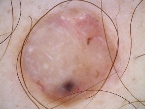
Polarized dermatoscopy view of nodular melanoma
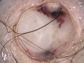
Non-polarized dermoscopy view of nodular melanoma, thickness 7.2 mm
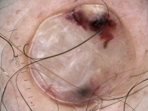
Polarized dermatoscopic view of nodular melanoma, thickness 7.2 mm
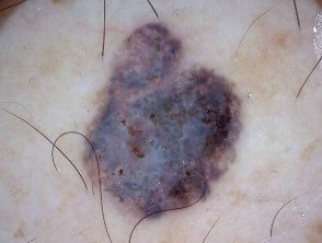
Non-polarized dermatoscopy of melanoma, 0.8 mm thick
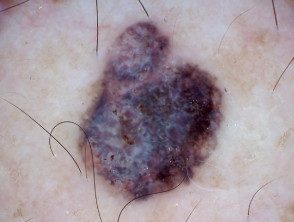
Polarized dermatoscopy of melanoma, thickness 0.8 mm
In which lesions are perpendicular white lines seen through the dermatoscope?
Perpendicular white lines can be seen in the following lesions:
- Pigmented and non-pigmented basal cell carcinoma
- Melanoma
- Spitz nevus
- Dermatofibroma (polarized white lines are often radial training instead of perpendicular)
- Scar tissue
- Benign lichenoid keratosis.
Perpendicular white lines in a variety of lesions.
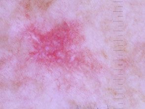
Superficial dermoscopy of basal cell carcinoma
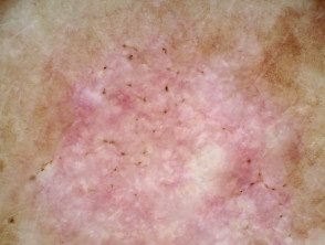
Superficial dermoscopy of basal cell carcinoma
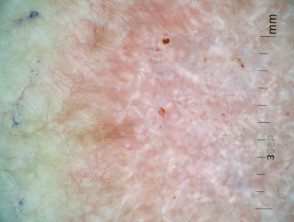
Superficial dermoscopy of basal cell carcinoma
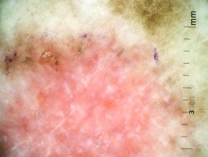
Superficial dermoscopy of basal cell carcinoma
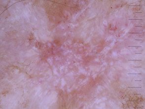
Basal cell nodular carcinoma dermoscopy
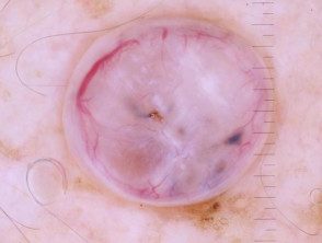
Polarized dermoscopy of a nodular basal cell carcinoma presenting as an exophytic polyp
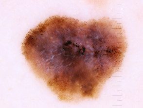
Invasive dermoscopy of melanoma, Breslow 0.4 mm within a melanoma in situ, with the presence of associated nevus
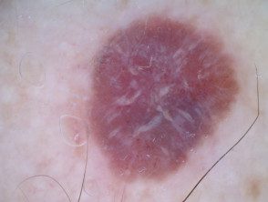
Amelanotic melanoma dermoscopy
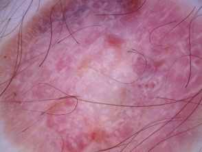
Amelanotic melanoma dermoscopy
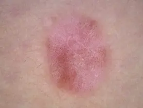
Spitz naevus dermatoscopy
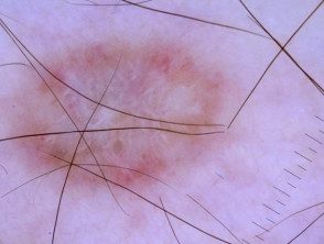
dermatofibroma dermoscopy
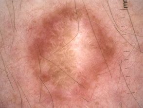
dermatofibroma dermoscopy
Which is the histological explanation of the perpendicular white lines?
Perpendicular white lines are believed to correlate histopathologically with altered collagen at dermis (fibrosis) The birefringent properties of collagen bundles cause rapid randomization of polarized light. This is the reason why collagen appears bright white and is more visible under polarized dermatoscopy. [3].
They also correlate with dermal invasion in melanoma cases [4]. However, as our illustrations show, they can also be seen in melanoma. in the place.