
What is it immunohistochemistry?
Immunohistochemistry (IHC) is considered an advanced form of histopathology. Immunohistochemistry is generally not used initially, but is added when routine / regular histological the test is insufficient to form a diagnosis.
IHC uses primary antibodies to tag a protein, then use a secondary antibody which is linked to the primary. In immunoperoxidase staining, an antibody binds to a enzyme, peroxidase, which catalyzes a reaction in which the protein specifically stains brown. IHC can also involve fluorescently labeled antibodies so that when viewed under a light microscope a certain pattern is observed from the emitted fluorescence.
The IHC pattern is considered diagnostic, which shows nuclear, membranous or cytoplasmic patterns. IHC is often used in situations where the presence or absence of certain proteins can form a basis for a diagnosis. It can also be used to distinguish between two different disease processes that might otherwise appear similar to pathologist.
How is immunohistochemistry performed?
The most common process for preparing immunohistochemical slides is as follows:
- Tissue fixation (in general, IHC stains are fixed with formalin)
- Embedding the tissue (in paraffin)
- Sectioning of tissues.
- Recovery (done with the application of heat or proteolytic enzymes)
- Assembly and dehydration.
- Cleaning and observation of the slides.
What are the advantages and disadvantages of immunohistochemistry?
The advantages of IHC include:
- Fresh or frozen tissue samples can be used for IHC.
- IHC is well established and readily available.
- The cost of IHC is relatively low
- It has a fast response time.
- Because no live infectious agents are involved, the risk to human health is minimal.
The disadvantages of IHC are as follows:
- IHC stains are not standardized throughout the world.
- Although the cost of the procedure is relatively inexpensive, the equipment required to perform IHC is expensive.
- Quantifying the results is difficult.
- IHC is subject to human error. Well-trained staff is paramount.
What are some examples of immunohistochemical stains?
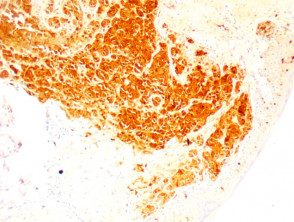
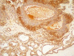
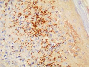
Hundreds of immunohistochemical stains are used to identify different tumors and other neoplasms. Only some of the IHC stains used in dermatology they are listed below.
| IHC Stain | Uses / Image Title |
|---|---|
| BCL2 | It is used to distinguish between basal cell carcinomas and trichoepitheliomas |
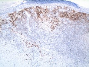
| CD3 | T cell marker; strongly positive in mycosis fungoides |
| CD4 | Helper T cell marker |
| CD8 | Suppressor T cell marker |
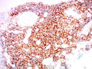
| CD20 | B cell marker |
| CD30 | Can be used in Hodgkin's diagnosis lymphoma and anaplastic Large cell lymphomas: Golgi apparatus and membranous staining. |
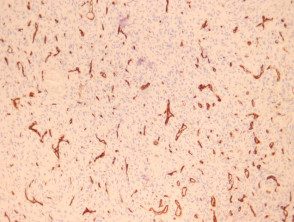
| CD31 | Helps to identify endothelial tumor |
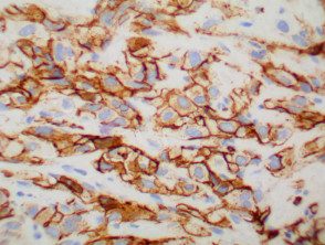
| CD34 | It distinguishes different endothelial tumors and is positive for dermatofibrosarcoma. |
| CD56 | Used in the diagnosis of non-Hodgkin's lymphomas, leukemias and small cell carcinomas |
| CD117 | Marker for KIT receiver and positive in several tumors, including mastocytosis |
| CDKN2A (p16) | Positive tumor suppressor marker in HPV-associated tumors, actinic keratosis and scaly cell carcinoma |
| CK (various) | Cytokeratins can be used to help distinguish benign since evil one attached tumors |
| CK 20 | Specific for Merkel cell carcinoma. May help identify adenocarcinomas of the gastrointestinal and reproductive system, as well as gastrointestinal epithelial tumors |
| Cytokeratin High Molecular Weight | It is used to detect ductal carcinomas, squamous cell carcinomas, and other epithelial neoplasms. |
| Desmin | Muscle marker |
| EMA | Used to identify eccrine neoplasms, Paget's disease and sebaceous carcinomas |
| Factor 13 | May help clinicians distinguish between dermatofibrosarcoma and dermatofibroma |
| HHV8 | Human herpes virus 8 |
| HMB 45 | Used to detect melanocytes, specially in melanoma but negative in desmoplastic melanoma |
| Melan-a | It can help identify melanocytic nevus cells and melanomas |
| PDL1 | Timed Death Ligand 1 |
| S-100 | It is used to mark tumors of melanocytes, both naevi and melanoma |
| SMA | Smooth muscle antigen |
| SOX-10 | Nuclear marker for melanocytic tumors |
| Treponema pallidum | Shows organisms in secondary syphilis |
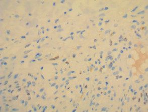
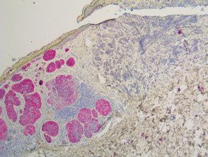
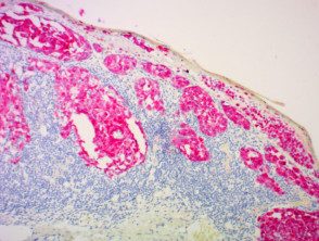
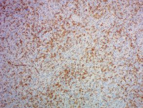
Immunohistochemical stains
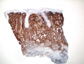
BCL2
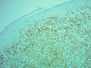
CD3

CD4
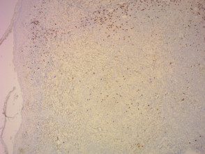
CD8
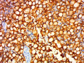
CD20
CD30
CD31
CD34
CD56
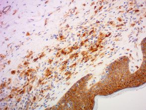
CD117
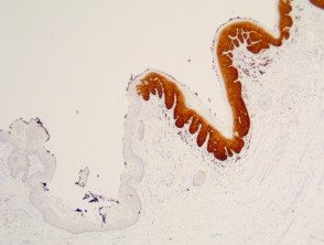
CDKN2A-P16
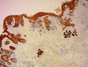
CK
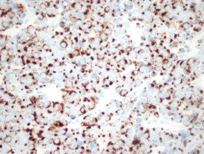
CK20
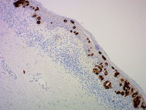
CK7
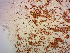
Desmin
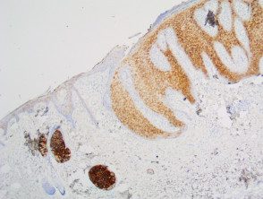
EMA
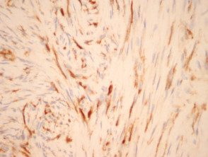
Factor 13a
HHV8
HMB45
Melan A
PDL1
S100
SMA
SOX10
Treponema pallidum
