Category

Aesthetic medicine blog

Marine animal bites: symptoms and treatment

starfish-bites-of-marine-animals-2081888-7201850-jpg-6039591

Conozca los síntomas y el tratamiento de las mordeduras de animales marinos como anémonas, corales, esponjas, erizos, pepinos y estrellas de mar.

En el mar encontramos invertebrados capaces de provocar lesiones cutáneas. Impactan a la población expuesta, pescadores, nadadores, surfistas, buceadores o bañistas.

Pueden ser muy frecuentes en la temporada de baño debido al aumento de la afluencia al mar, y provocar molestias y daños en la piel, a pesar de todo, las reacciones graves son muy raras.

En este artículo abordamos las dermatosis provocadas por mordeduras de animales marinos como erizos de mar, estrellas, esponjas o anémonas. Dermatología marítima para conocimiento de los que acuden ocasionalmente y mejora de los habituales.

Debemos recordar que primera medida En los primeros auxilios y tratamiento de picaduras en el mar, se trata de sacar a la víctima del agua, verificar que esté estable, dejarla en reposo y tranquilizarla.

7 mordeduras de animales marinos que debes saber

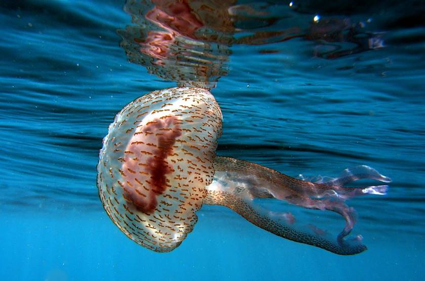
1 Dermatitis por anémonas o fideos de mar

Anémonas

• La Anémonas de mar Son animales marinos que se adhieren a las rocas o la arena.

• Las picaduras de anémona pueden causar dermatitis en pescadores de esponjas, bañistas, buceadores y personas que recolectan moluscos de huecos en las rocas.

• Sus tentáculos producen reacciones de urticaria con apariencia de ardor, enrojecimiento y picor cuando entran en contacto con la piel.

• El tratamiento se trata de retirar los tentáculos y aplicar vinagre en la zona.

• Se resuelve lentamente en 1-2 semanas.

2 Erupción del bañista

Edwarsiella lineata - Mordeduras de animales marinos

• Fue descrito por primera vez en 1949 en bañistas de Florida. Las áreas donde ocurre con mayor frecuencia son Florida, el Caribe, Bermudas y la costa noroeste de los Estados Unidos.

• Es causada por las larvas de una anémona (Edwarsiella lineata) que inyectan la toxina de forma directa en la piel.

• Suele producir lesiones en la zona del traje de baño y donde la ropa o los accesorios entran en contacto con la piel, reteniendo la larva en ella.

• Comienza con mucha picazón que se transforma en vesículas, pápulas y reacción de urticaria en la zona afectada que cede en una semana.

• El 10% de los pacientes pueden presentar síntomas sistémicos como malestar, fiebre o náuseas.

• El tratamiento es la retirada del bañador lo antes viable. Se pueden utilizar corticosteroides tópicos para reducir los síntomas posteriores.

3 Lesiones cutáneas por corales

3.1 Dermatitis de coral, coral del mar rojo o coral de fuego

• La dermatitis de coral en general es bastante inofensiva excepto en el caso del coral de fuego. Este coral está en Japón y en el Pacífico.

• Causada por Dendornephtya nipponica, una especie animal muy simple que parasita las langostas y libera nematocistos (larvas) que irritan la piel.

• Afecta a los pescadores de septiembre a abril en forma de urticaria, ampollas, rinitis y conjuntivitis. Además puede afectar a los bañistas.

• Las mordeduras de animales marinos, como los corales, deben enjuagarse con agua de mar. Al mismo tiempo, se deben borrar los nematocistos que no se hayan descargado.

• El área de la picadura se puede cubrir con una compresa de vinagre o alcohol diluido al 40-70% durante 15 a 30 minutos hasta que se alivie el dolor. Además se pueden usar compresas de agua de mar lo más calientes viable para inactivar nematocistos. Después, se puede aplicar una crema con corticosteroides.

3.2 Desgarros o heridas de coral y su tratamiento:

• Son producidos por sus exoesqueletos afilados y tardan un tiempo notable en sanar. Además disponen un mayor riesgo de infección.

• El tratamiento se trata de una limpieza profusa de la herida con agua y jabón. Dependiendo del tipo de herida, se puede considerar coserla y además se debe considerar la vacuna contra el tétanos.

4 erizos de mar

Erizos de mar - Picaduras de animales marinos

• Viven en el suelo, cerca de las costas de todo el mundo.

• Las picaduras de animales marinos como los erizos suelen causar daños en el mar.

• Contienen púas afiladas que a veces pueden contener veneno, lo que además produce una reacción local.

• Las púas se clavan en la piel y las más pequeñas pueden tener acceso.

• El tratamiento se trata de borrar las espinas que se puedan hacer y lavar la zona con alcohol o antisépticos:

– Las espinas son muy frágiles y algunas rompen la piel y no se pueden borrar.

– No se deben realizar más intentos fuera del ambiente sanitario y los que queden introducidos en la piel se reabsorberán con el tiempo.

– En las púas grandes, se puede usar una radiografía para confirmar que no afecta a las articulaciones ni a los tendones.

– Se debe considerar la vacuna contra el tétanos y de forma general no necesita antibióticos preventivos.

– Las púas de erizo de color marrón oscuro pueden dejar la piel pigmentada. Esta pigmentación se desvanece con el tiempo.
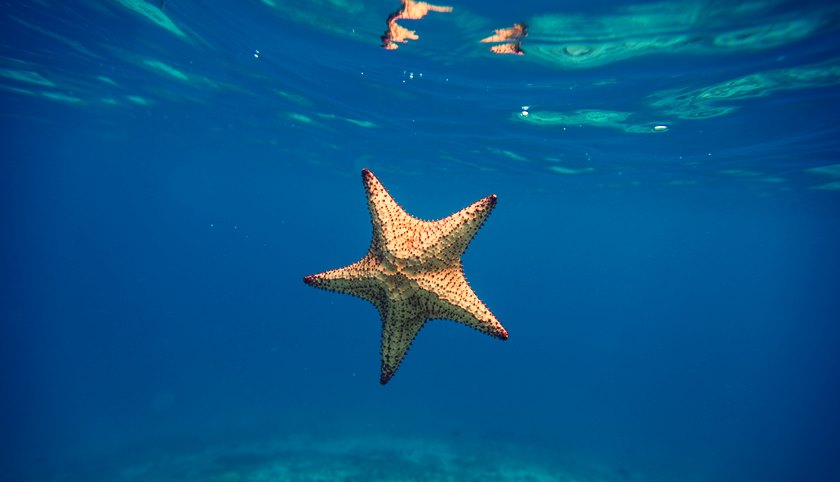
5 estrellas de mar

Estrella de mar

• No suelen dañar a los bañistas.

• A pesar de todo, el contacto con la estrella de mar “Corona de espinas” localizada en la Gran Barrera de Coral puede ser muy dañino.

• Produce diversos heridas punzantes con sus espinas que son capaces de penetrar el neopreno.

• El tratamiento es equivalente al de las picaduras de animales marinos como el erizo de mar. Las espinas deben eliminarse siempre y cuando sea viable y se debe considerar la vacunación contra el tétanos.

6 pepinos de mar

Pepino de mar - Picadura de animales marinos

• Viven en el fondo del mar y producen una toxina en su superficie.

• El contacto puede causar dermatitis irritativa y conjuntivitis.

• El tratamiento incluye el lavado inmediato con agua tibia con o sin jabón, vinagre o alcohol.

7 Lesiones cutáneas por esponjas

Esponjas de mar - Mordeduras de animales marinos

7.1 Dermatitis por espículas esponjosas

• Las esponjas viven en el fondo del mar y algunas pueden tener espículas capaces de causar dermatitis irritativa o abrasiones menores.

• La reacción aparece en minutos.

• El tratamiento se trata de la eliminación de las espículas con cinta adhesiva o esparadrapo. Algunos autores recomiendan aplicar arena de la playa de la zona para inactivar las espículas.

7.2 Dermatitis por esponja tóxica

• Descrito en al menos 13 especies, ninguna de ellas se encuentra en nuestras costas.

• Se produce al tener acceso en contacto con un veneno que disponen en su superficie.

• Produce síntomas locales graves en 24 horas en forma de hinchazón, ampollas y dolor.

• El tratamiento se trata de lavar la zona, aplicar vinagre y poner compresas frías.

References

1 Tlougan BE, Podjasek JO, Adams B. Dermatosis de deportes acuáticos. Parte 2. En el agua: dermatosis de agua salada. Int J Dermatol 2010; 49: 994-1002.

2 Reckziegel GC, Dourado FS, Garrote-Neto, et al. Lesiones causadas por animales acuáticos en Brasil: un análisis de los datos presentes en el sistema de información de enfermedades notificables. Rev Soc Bras Med Trop 2015; 48: 460-467.

Te invito a compartir este post para que otras personas conozcan los síntomas y el tratamiento de las mordeduras de animales marinos como Anémonas, Corales, Esponjas, Erizos, Pepinos y Estrellas de Mar.

You may also be interested in:

Treatment of jellyfish stings and strategies to avoid them

jellyfish-sting-treatment-6395877-9547371-jpg-7357170

Picaduras de medusa, ¿se tratan con amoniaco, vinagre o pipí? ¿Cómo se tratan las picaduras en el mar? En este artículo, repaso la literatura y mi experiencia para conocer el tratamiento de las picaduras de medusa..

En los últimos años el aumento del crecimiento de las medusas está causando una mayor relevancia de su picadura. Este aumento puede deberse al cambio climático, la sobrepesca y la alteración de los hábitats marinos. Hay más de 10,000 especies de medusas de las cuales el 1% generan daño a los humanos.

Especies de medusas más reconocidas

Las especies más reconocidas son: Escifozoos o “medusa verdadera”, Cubozoa (“Medusa de caja”) y Hidrozoos (hidropólipos e hidromedusas o falsas medusas).

El cuidado de las picaduras de medusas es la actividad sanitaria más usual en las playas del Mediterráneo, y se estima que cada verano se producen unas 25.000 picaduras solo en la costa catalana (1).

the carabela portuguesa o fragata (Physalia physalis) nos suena familiar a todos, es un Hydrozoo o una falsa medusa. Está en el mar abierto de todas las aguas templadas del mundo, es un organismo colonial que forma un auténtico velero. Es capaz de producir reacciones severas pero su presencia en nuestras cosas es rara. Como veremos más adelante, el tratamiento de su picadura difiere significativamente del tratamiento de la picadura de sus “primas” las medusas.

Hombre de guerra portugués - Tratamiento de picaduras de medusas

Tratamiento de picadura de medusa: ¿Qué dicen los estudios científicos?

Hay mucha información en la literatura acerca de cómo tratar las picaduras de medusa y, a veces, es contradictoria, injustificada y poco clara. Hay poca evidencia basada en la evidencia y se aceptan recomendaciones anecdóticas, que en ocasiones pueden inclusive agravar los síntomas de la picadura.

Si revisamos la literatura, podemos resumir varias ideas clave con respecto al tratamiento:

• Hay un amplia variabilidad en la picadura según la especie que lo provoca: la picadura depende de la especie, la zona geográfica, la composición del veneno de la medusa, el lugar de contacto, etc. Hay especies no venenosas, cuya toxina no es capaz de penetrar en la dermis. Y además se sabe que no todos los tentáculos son capaces de provocar una reacción (2).

• Existe variabilidad según cada personaNadie es resistente a la picadura de una medusa, pero se sabe que afecta a cada persona de manera distinto. La mayoría de las reacciones se limitan a la piel de la zona de contacto, en forma de urticaria y eritema en la piel (enrojecimiento) que producen picor y dolor de distinta intensidad y duración. A pesar de todo, se han descrito reacciones sistémicas más graves y muy raras con angioedema y anafilaxia.

• La reacción que producen se debe a acción irritante directa de la toxina de las medusas en la piel o por estimulación del respuesta inmune de linfocitos. Esta respuesta inmune es celular de linfocitos T y humoral de linfocitos B. Para que se produzcan reacciones alérgicas, con agioedema o anafilaxia, hace falta haber tenido una exposición previa al veneno, dicho de otra forma, haber sido picado antes (1).

Primeros auxilios y tratamiento de picaduras de medusas

Tratamiento de picaduras de medusas

¿Qué debemos hacer ante una picadura de medusa?

Un interesante post resume el protocolo de actuación ante una picadura de medusa en Europa según la evidencia publicada en estudios científicos, es el siguiente (2,3):

1 Rescate al paciente y asegúrese de que esté seguro y estable.

two Asegúrese de que la víctima permanezca relajada y sin moverse para evitar la circulación del veneno.

3 Administra un analgésico oral.

4 Limpiar los tentáculos con agua de mar, NO con agua del grifo.

5 SOLO para medusas verdaderas y medusas de caja: remoje o empape la piel afectada con vinagre (4-6% de ácido acético) durante al menos 30 segundos. Parece que esta medida puede ser controvertida en las picaduras del buque de guerra portugués que recordamos es una hidromedusa o falsa medusa (4).

6 Si quedan tentáculos adheridos a la piel, quítelos con las manos enguantadas.

7 Para ortigas de mar: aplicar una solución de bicarbonato de sodio (50% de bicarbonato de sodio y 50% de agua de mar durante varios minutos y enjuagar con agua de mar. O rociar bicarbonato de forma directa sobre los tentáculos).

8 Si ninguno de los anteriores se encuentra disponible, el área afectada se puede sumergir en agua caliente al 42-45% continuamente hasta que se elimine el dolor o el área se pueda enjuagar con agua de mar.

9 NO use agua dulce, licor o alcohol para desactivar los tentáculos de la piel, dado que todos pueden causar una descarga masiva de nematocistos in some cases.

10 Hace falta acudir a urgencias si el paciente comienza con síntomas sistémicos (dolor abdominal, hipotensión, mareos, náuseas, vómitos, enrojecimiento generalizado de la piel) o el dolor no cede.

once El tratamiento posterior de los síntomas se realiza con antihistamínicos para la picazón, corticosteroides tópicos para disminuir la inflamación. En casos más graves, se pueden prescribir corticosteroides orales o intramusculares.

12 Además encontramos referencias menores en la literatura a la oportunidad de usar agua fría o compresas frías (4).

13 NO existe evidencia en la literatura del uso de coca-cola, alcohol o pipí en picaduras de medusas (4).

¿Cómo podemos prevenir las picaduras de medusas?

Prevención de picaduras de medusas.

1 Nadar en playas controladas y con primeros auxilios o centros de atención adecuados.

two Evite nadar en aguas con medusas, sobre todo posteriormente de una tormenta, dado que las picaduras pueden ser causadas por los restos de tentáculos dañados que flotan en el agua.

3 Cuidado con las medusas aparentemente muertas o que se encuentran en la arena de la playa.

4 Use ropa protectora si practica esnórquel, bucea o nada en aguas abiertas.

5 Use protector solar con repelentes para medusas.

Limitaciones de los estudios sobre el tratamiento de las picaduras de medusas

Estas recomendaciones son generales y pueden variar en eficacia según el tipo de medusa y la reacción de cada paciente.

Cabe destacar que, aunque estas medidas recomendadas son las que más evidencia disponen en la literatura no hay grandes estudios o ensayos clínicos eso aumentaría la calidad de la evidencia para los tratamientos para las picaduras de medusas.

La base de datos Cochrane de revisiones sistemáticas (Revisiones sistemáticas de la base de datos Cochrane), que es la principal revista y base de datos de revisiones sistemáticas en salud, dicho de otra forma, un culto para los médicos, afirma. Parece lógico que no los haya en otros lugares. Y orinar no funciona.

References

1 Nogué S, Velasco V, Marambio M, Lopez L. Picaduras de medusas y otros animales marinos venenosos: su impacto en la actividad de los socorristas en la playa. Emergencias 2017; 29: 426-434-

two Montgomery L, Seys J, Mees J. Para orinar o no orinar: una revisión sobre el envenenamiento y el tratamiento en especies de medusas europeas. Drogas marinas 2016; 14: 127.

3 Li L, Mc Gee RG, Isbister G, Webster AC. Las intervenciones o los síntomas y signos dan lugar a picaduras de medusa. Syst Rev de la base de datos Cochrane 2013; 9 de diciembre.

4 Haddad V. Dermatología ambiental: manifestaciones cutáneas de lesiones provocadas por animales acuáticos invertebrados. Un sujetador Dermatol 2013; 88: 496-506.

Te invito a compartir este post para que otras personas tengan un conocimiento adecuado sobre el tratamiento de las picaduras de medusa y aprendan algunas estrategias para evitarlas.

Si deseas conocer el tratamiento de las mordeduras de otros animales marinos (como Anémonas, Corales, Esponjas, Erizos, Pepinos y Estrellas de Mar): Click here.

You may also be interested in:

6 foods you can eat to improve your vitiligo

foods-improve-vitiligo-8120968-9531742-jpg-9665966

Learn about the 6 foods you can eat to improve your vitiligo according to scientific literature.

Vitiligo is a dermatosis that causes white spots on the skin as a result of the destruction of pigment-producing cells (melanocytes).

It affects approximately 1% of the population and even when it produces nothing more than spots on the skin, patients are affected by their image. The cause of vitiligo is not known, but it is known that patients with vitiligo suffer from oxidative stress on your skin that destroys melanocytes.

In this post, I review how some foods can help reduce that stress on the skin and help traditional treatments for vitiligo protect melanocytes. In other words, I am going to tell you several foods that you can eat to improve your vitiligo.

What is vitiligo and how is it treated?

Vitiligo is a dermatosis that affects approximately 1% of the population and produces white spots on the skin. The disfigurement caused by vitiligo affects the quality of life of patients.

The treatment of vitiligo requires the combination of topical treatments in the form of creams and phototherapy (light that imitates the sun on the skin) in more extensive cases to recolor (re-pigment) the spots.

In my experience as a dermatologist, vitiligo treatment is long, requiring a minimum of 6 to 12 months and a lot of patience. Vitiligo requires long-term treatment and is not always effective, so the doctor-patient relationship should be maintained as much as possible. It is essential that the patient feels comfortable and that we honestly explain the treatment and their expectations.

On the other hand, the melanocyte, like the patient, must also be treated with care. And when it appears, in other words, when the patient begins to re-pigment, it is essential that it does not disappear again. This is why, in my opinion, it is essential to promote an environment without oxidative stress in the skin appropriate for the melanocyte.

In more scientific terms, elevated markers of oxidative damage are found in the epidermis and blood of vitiligo patients, producing ROS (oxygen free radicals) and hydrogen peroxide (H2O2) (1). There are several studies that suggest that oxidative stress is crucial in the genesis of vitiligo, causing inflammation, autoimmunity and death of melanocytes in the skin (2).

Discover the 6 foods you can eat to improve your vitiligo

Immunonutrition attempts to modulate the immune system through the supply of nutrients. It is an emerging and promising discipline. Here you can visit the web portal of the International Society for Immunonutrition, ISIS (@ISImmunoNutr).

There are no studies or clinical trials in the reviewed literature that demonstrate how diet affects patients with vitiligo, thus, dietary recommendations are made approximately, looking for antioxidant and anti-inflammatory foods.

And what are the functional foods in vilitigo?

1. Green tea

It has been shown in in vitro (laboratory) and animal studies to protect melanocytes from oxidative stress (3).

2. Indian Gooseberry or Amla Fruit (Phyllanthus emblica)

A powerful oral antioxidant that also enhances the antioxidant action of vitamin C and E. In a group of patients with vitiligo treated with phototherapy, those who were super-supplemented with Amla and vitamins C, E and carotenes repigmented more and faster (4 ). Therefore, it is another of the foods that you can eat to improve your vitiligo.

3. Gingko Biloba

A classic. Its benefits as an antioxidant have been proven in various studies and it is one of the most studied medicinal herbs. In doses of 240 mg per day it is beneficial without producing side effects. In a 12-week clinical trial, Gingko biloba 60 mg daily in patients with vitiligo decreased the affected area (Vitiligo Area Score Index, VASI and European Vitiligo Task Force, VETF) (5).

4. Polypodium leucotomos

It is an extract from the fern grown in South America and is taken orally in doses of 250-720 mg per day. In Spain it is sold in pharmacies, patented under the name Fernblock. It has no side effects except for slight gastric discomfort. It has been shown in two clinical trials to increase repigmentation opportunities and repigmentation area by approximately 50% in vitiligo patients treated with phototherapy (6). As a dermatologist who is an expert in the treatment of vitiligo with phototherapy, I use it on all my patients.

5. Kelina (Ammi visnaga or biznaga extract)

It is an extract from an herb that grows wild in the Mediterranean. At a dose of 100 mg per day, it has been shown in two clinical trials combined with UVA, to increase the chances of repigmentation by approximately 50% on average. It can cause nausea and elevated transaminases (7). Additionally, 3% can be used as a topical cream combined with sun or UVA exposure, in a phototherapy modality called KUVA. As a vitiligo dermatologist, I use it on my vitiligo patients in the summer to avoid trips to the clinic for phototherapy.

6. turmeric or turmeric

Another classic to highlight among the foods you can eat to improve your vitiligo. It is capable of reducing the production of ROS in in vitro studies in the skin of patients with vitiligo. Its topical application combined with phototherapy appears to slightly increase the repigmentation rate of vitiligo spots.

Finally

In summary, functional nutrition along with an appropriate diet in vitiligo combined with medical treatments can be another option in the management of patients. Amla Fruit, Gingo Biloba, Kelina and Polypodium have been studied in patients with vitiligo improving the solution to phototherapy, and the latter is the most used in our country.

References

1. Mansuri MS, Jadeja S, Singh M, et al. Catalase gene promoter and untranslated region variants lead to altered gene expression and enzymatic activity in vitiligo. Br J Dermatol 2017; 177: 1590-1600.

2. Marie J, Kovacs D, Pain C. Inflammasome activation and progression of vitiligo/nonsegmental vitiligo. Br J Dermatol 2014; 170: 816-823.

3. Ning W, Wang S, Liu D et al. (2016). Potent effects of peracetylated-epigallocatechin-3-gallate against hydrogen peroxide-induced damage in human epidermal melanocytes through attenuation of oxidative stress and apoptosis. Experimental Clinical Dermatology2016; 41: 616–624.

4. Colucci R, Dragoni, F, Conti R, et al. Evaluation of an oral supplement containing Phyllanthus emblica fruit extracts, vitamin E and carotenoids in the treatment of vitiligo. Dermatological therapy 2015; 28:17-21.

5. Szczurko O, Shear N, Taddio A. et al. Ginkgo biloba for the treatment of vitiligo vulgaris: an open-label pilot clinical trial. BMC Complementary and Alternative Medicine 2011; eleven; twenty-one.

6. Middelkamp-Hup, MA, Bos, JD, Rius-Diaz, F., González, S. and Westerhof, W. (2007). Treatment of vitiligo vulgaris with narrow-band UVB and oral extract of Polypodium leucotomos: a randomized, double-blind, placebo-controlled study. Journal of the European Academy of Dermatology and Venereology, 21 (7), 942–950.

7. Hofer, A, Kerl H, Wolf P. Long-term results in the treatment of vitiligo with oral khellin plus UVA. European Journal of Dermatology 2001; eleven; 225-229.

8. Asawanonda, P, Klahan S. Tetrahydrocurcuminoid cream plus targeted narrow-band UVB phototherapy for vitiligo: a preliminary randomized controlled study. Photomedicine and Laser Surgery 2010; 28; 679-684.

Nutrition and diet tips to improve psoriasis

diet-improve-psoriasis-8933142-4095571-jpg-7596026

Psoriasis is a very common dermatosis that affects the 2% of the general population with variable severity. Psoriasis is of multifactorial origin, in other words, caused by various factors that act on a genetic predisposition and cause skin inflammation.

Among the risk factors described there is accumulating evidence that nutrition plays a role. This post summarizes the scientific studies made public about nutritional and dietary strategies to improve psoriasis, the benefits of some dietary supplements and probiotics.

What is psoriasis and how is it treated?

The psoriasis It is a very common dermatosis which affects up to 2% of the population worldwide. Its treatment depends on the severity and includes topical treatments in the form of creams, systemic pills and phototherapy.

However, on repeated occasions the patient asks us what to eat, if there is any guideline in his diet to improve the psoriasis he suffers from.

On other occasions, the patient demands a more comprehensive approach, towards modern medicine, in which we treat their psoriasis in a more global way. The link between psoriasis and nutrition is widely studied in scientific literature; we can summarize it in three fundamental points: diet as a strategy, the use of nutritional supplements and probiotics.

Nutrition and diet strategies to improve psoriasis: What should I eat?

Little, undoubtedly. The obesity is a risk factor very important in psoriasis, and it is bidirectional, in other words, obesity worsens psoriasis and psoriasis promotes obesity (1). Obese patients have twice the risk of having psoriasis than those who are not obese.

Several lines of research suggest that adipocytes and their inflammatory molecules, adipokines, play a role in skin inflammation and in the hypothalamus, the main hunger regulatory center. Obesity releases a multitude of mediators into the blood that produce a situation inflammatory in the body. Leptin, a major intake-regulating hormone, is elevated in patients with psoriasis compared to healthy controls (2).

Obesity also has many implications for psoriasis treatments, as follows:

  • When the patient loses weight, the response to treatments is better or they even stop needing them.
  • Obesity increases the risk of side effects from systemic treatments (pills or injections).

The hypocaloric diet (800-1200 Kcal/day) combined with aerobic exercise at least 3 times a week for 30 minutes has been shown to improve psoriasis in a few weeks in many studies (3,4).

Components of the diet to improve psoriasis (3,5):

Diet rich in fish oils (omega-3 polyunsaturated fatty acids, PUFA), present in oily fish. They are the ones with the most evidence, due to their ability to reduce arachidonic acid and promote an anti-inflammatory environment.

Diet rich in monounsaturated fatty acids (MUFA) present in extra virgin olive oil, rich in oleic acid. Low MUFA intake worsens psoriasis and may cause it to progress. This study is in line with several others published about MUFAs and their anti-inflammatory capacity.

Regarding the type of global diet, Mediterranean diet takes the cake (3,4,5). We have plenty of scientific evidence that eating patterns rich in fruits, vegetables and fish are associated with a reduction in global markers of inflammation. In this situation it seems logical as it has been shown in numerous studies that the Mediterranean Diet improves psoriasis.

exist others publications that address different diet options to improve psoriasis, but with more limited scientific evidence, so their recommendation is not clear today (3):

  • Gluten-free diet: it has been clearly shown to be beneficial in patients with celiac disease, also latent, and psoriasis (5).
  • Vegetarian diet.
  • Low carbohydrate diet.

Beneficial food supplements to treat psoriasis

Vitamin D. has been shown to have an anti-inflammatory role through the regulation of regulatory T lymphocytes. It should undoubtedly be supplemented in cases of deficiency since its deficiency can worsen psoriasis (3,4). However, its supplementation in patients with normal levels of vitamin D has not shown clear benefit (5,6). As a dermatologist who is an expert in psoriasis and phototherapy in Madrid, I do use it as a supplement in the patients I treat for psoriasis with phototherapy, due to its ability to protect the skin from the sun.

Fish oil and omega 3 supplements. They have been shown in numerous clinical trials to be beneficial in patients with psoriasis and other dermatoses (6). They must be maintained for long periods of up to 6 months to achieve effective benefit in psoriasis.

Supplements antioxidants In general, with vitamins (Vitamins A, E, C, folic acid, group B vitamins), trace elements such as Copper, Zinc, Iron, Magnesium and Selenium, coenzyme Q10 for its antioxidant capacity. They have demonstrated their positive effects in important studies (7,9).

  • Zinc Supplements: Zinc is a trace element that is capable of regulating the immune response. The evidence of supplementing zinc in psoriasis is controversial, thus we find studies in which they have shown improvement and others have not (7,8).
  • Selenium supplements: This is a trace element with antiproliferative capacity. Low selenium levels can worsen psoriasis (9).

Complementary medicinal plants or as an option to improve psoriasis

Medicinal plants are increasingly used and we have various studies and clinical trials published in quality scientific literature in which they demonstrate effectiveness in psoriasis. Such is the case of Dunaniella. bardawil, TwHF( Tripterigium wilfordii), Azadirachta indica (Neem tree) or Turmeric. Longa. However, at the current time we do not have guides for its use.

Polypodium Leucotomos

It is an extract of a fern (patented under the name of Fernblock) which has been shown to reduce the response of CD4 lymphocytes when taken orally. Studies of this compound were carried out in Spain years ago and demonstrated a mild to moderate benefit in half of the treated patients (10).

It is available sold in pharmacies and until a few years ago it was financed by the National Health System. We can say that it is the only plant available in Western medicine for the treatment of psoriasis.

This compound is also of special interest as an antioxidant and oral sunscreen.

Probiotics, prebiotics and synbiotics that help against psoriasis

Probiotics, prebiotics and synbiotics are other components to consider along with herbal remedies and dietary advice to improve psoriasis.

As a brief introduction, I will first break down what each of them is:

  • Probiotic: a microorganism that, taken, produces health benefits.
  • Prebiotic: a substance that is ingested promotes the growth of some bacteria in the colon that are beneficial for health
  • Symbiotic: It is the mixture of the previous two

Bifidobacterium infantis

To date we only have one study of a probiotic in psoriasis: it is Bifidobacterium infantis 356624 (yes, probiotics have a serial number along with the name of the bacteria).

This bacteria was ingested for 8 weeks by psoriasis patients who were compared to psoriasis patients who did not take it. It was shown that it is capable of reducing the classic markers of inflammation in psoriasis such as TNF-alpha and the interleukin 6 or C-reactive protein; However, it did not translate into a clear benefit for skin lesions, so, even though it seems promising, B. infantis more studies are needed.

Conclusion

Psoriasis benefits from antioxidant rich diet (fruits, vegetables), fish, especially blue, and olive oil in a controlled calorie intake. Approximately, Mediterranean diet.

Furthermore we can add blue fish and antioxidant supplements diet to improve psoriasis. Supplementing with vitamin D or selenium makes sense in patients with a deficiency, since this situation can worsen psoriasis.

There is evidence of improvement in psoriasis with some oral plants (herbal medicine), but we currently have no guidelines for its use. The extract of Polypodium leucotomos It is safe orally and can improve in about half of patients. And at probiotics they have abandoned.

Bibliography

1. Carrascosa JM, Rocamora V, Fernandez-Torres RM et al. Obesity and psoriasis: inflammatory nature of obesity, link between psoriasis and obesity and therapeutic implications. Actas Dermosifiliofr 2014; 105: 31-44.

2. Zhu KJ, Zhang C, Li M. Leptin levels in patients with psoriasis: a meta-analysis. Clin Exp Dermatol 2014; 222: 113-127.

3. Barrea L, Nappi F, Di Somma C, et al. Environmental risk factors in psoriasis: the nutritionist's point of view. Int J Environ Res Public Health 2016; 13: 743-755.

4. Zuccotti E, Oliveri M, Girometa C, et al. Nutritional strategies for psoriasis: current scientific evidence in clinical trials. Eur Rev Med Pharmacol Sci 2018; 22: 8537-8551.

5. Ford A, Siegal M, Bagel J. A dietary recommendations for adults with psoriasis or psoriatic arthritis from the medical board of the national psoriasis foundation. JAMA Dermatol 2018; 154: 934-950.

6. Millsop JW, Bathia BK, Debbaneh M, et al. Diet and psoriasis, part III: role of nutritional supplements. J Am Acad Dermatol 2014; 71: 561-569.

7. Smith N, Weyman A, Tausk et al. Complementary and alternative medicine for psoriasis: a qualitative review of the clinical trial literature. J Am Acad Dermatol 2009; 61: 841-856.

8. Wacemicz M, Socha K, Soroczyska J, et al. Selenium, zinc, copeer concentration, Cu/Zn binding, total antioxidant status and serum c-reactive protein of psoriasis patients treated with narrowband ultraviolet B phototherapy: a case-control study. J Trace Elem Med Biol 2017; 44: 109-114.

9. Kharaeva Z, Giostova E, De Luca et al. Clinical and biochemical effects of supplementation with coenzyme Q10, vitamin E and selenium in patients with psoriasis. Nutrition 2009; 25: 295-302.

10. Padilla HC, Laínez H, Pacheco JA. A new agent (hydrophilic fraction of polypodium leucotomos) for the treatment of psoriasis. Int J Dermatol 1974; 13: 276-282.

11. Groeger D, O'Mahoni L, Murphy EF, et al. Bifidobacterium infantis 35624 modulates host inflammatory processes beyond the intestine. Gut Microbes 2013; 4: 325-339.

What is the best clothing for atopic dermatitis?

best-clothes-atopic-dermatitis-3964479-5772285-jpg-9817079

It is common for patients or parents of children to ask us what is the best clothing for atopic dermatitis.

Classically, dermatologists recommend cotton clothing and, on the other hand, patients often report intolerance to wool. The scientific literature can to surprise regarding this recommendation.

A usual question in the consultation

Patients with moderate-severe atopic dermatitis often consult us about factors that can aggravate or improve their dermatitis.

A common question in everyday dermatology is whether there are more suitable garments. In a traditional way we suggest cotton or natural fiber clothing, which is breathable and does not increase itching or cause skin irritation.

I also recommend cotton pajamas to go to bed after bathing and moisturizing the skin, with or without corticosteroid cream, I find them very practical to make a slight occlusion of the applied cream and facilitate its penetration.

But if we review the quality scientific literature, what studies do we find of clothing in atopic dermatitis?

What the science says about the best clothes for atopic dermatitis

I find the Study DESSINE published in the magazine of the British Association of Dermatology in 2017 (1), one of the most impressive in dermatology.

In this work, they study two groups of pediatric patients with moderate-severe atopic dermatitis. One group of patients wears cotton clothes and another group is dressed in superfine sheep's wool clothes.

They study the two groups and measure in them the severity of atopic dermatitis with normalized indices, the SCORE, the atopic dermatitis severity index (ADSI) and the quality of life index in children with dermatitis.

Surprisingly, they find that patients using wool clothing his dermatitis improves throughout follow-up.

Despite limited evidence, dermatologists have considered wool an irritating fabric that should be avoided in patients with atopic dermatitis.

At the same time, in Hanifin and Rajkapara criteria the diagnosis of atopic dermatitis, one of which is included is wool intolerance.

Wool fibers are composed of keratin, have insulating capacity, have a greater composition of water than cotton fibers, are thermoregulatory and transport moisture.

These qualities of wool can be beneficial in patients with atopic dermatitis and have been shown in children and adults (2). This study makes us reflect on the traditional recommendation to wear cotton clothing.

Other studies

In addition there are other interesting studies on textiles in atopic dermatitis in the literature:

  • Textiles with an antimicrobial effect, coated with antiseptic substances (3), have shown to be beneficial, despite everything there are still studies to recommend their use on a regular basis and they are not included in the Management Guidelines (4).
  • The CLOTHES study evaluates therapeutic silk garments, without showing improvement with them in children with atopic dermatitis (5).
  • Another recent study finds benefits in clothing made from cellulose fibers enriched with silver-containing algae (6).

Conclusions on what is the best clothing for atopic dermatitis

We can conclude that the classic recommendation to use cotton fabrics in atopic dermatitis is mainly based on the sensation subjective of the patient, who feels more comfortable, but fine wool clothing may be beneficial in these patients.

Other tissues and components are being studied without there being any specific recommendation in the management guidelines at the moment. studies are missing to establish the best clothing for atopic dermatitis on a solid foundation.

References

1 His JC, Dailey R, Zallmann, et al. Determination of the effects of superfine sheep's wool on childhood eczema (DESSINE): a randomized pediatric crossover study. Br J Dermatol 2017; 177: 125-133.

two Griplas L. Changing perceptions of wool. Sydney: Woolmark, 2012. Available at:. [8 May 2014].

3 Lopes C, Soares J, Tavaria F, et al. Chitosan-coated textiles may improve the severity of atopic dermatitis by modulating the staphylococcal profile of the skin: a randomized controlled trial. Pls One 2015; 30: 1-14.

4 Lopes C, Silva D, Delgado L, Correia O, Moreira A. Functional textiles for atopic dermatitis: a systemic review and meta-analysis. Pediatr Allergy Immunol. 2013; 24(6):603-13.

5 Thomas K, Bradshaw L, Sach T, et al. Randomized controlled trial of therapeutic silk garments for the treatment of atopic eczema in children: the CLOTHES trial. Health Technology Assessment 2017; 9:16 p.m.

6 Portela Araujo C, Gomes J, Paula Vieira F, Fernandes JC, Brito C. An initiative for the use of novel silver algae cotton fibers in the treatment of atopic dermatitis. Cutan Ocul Toxicol, 2013; 32: 268-274.